Review
. 2021 May 11;54(5):859-874.
doi: 10.1016/j.immuni.2021.01.012. Epub 2021 Apr 10.

Chemokines and the immune response to cancer

Affiliations
Review

Chemokines and the immune response to cancer

Aleksandra J Ozga et al. Immunity. .

Abstract

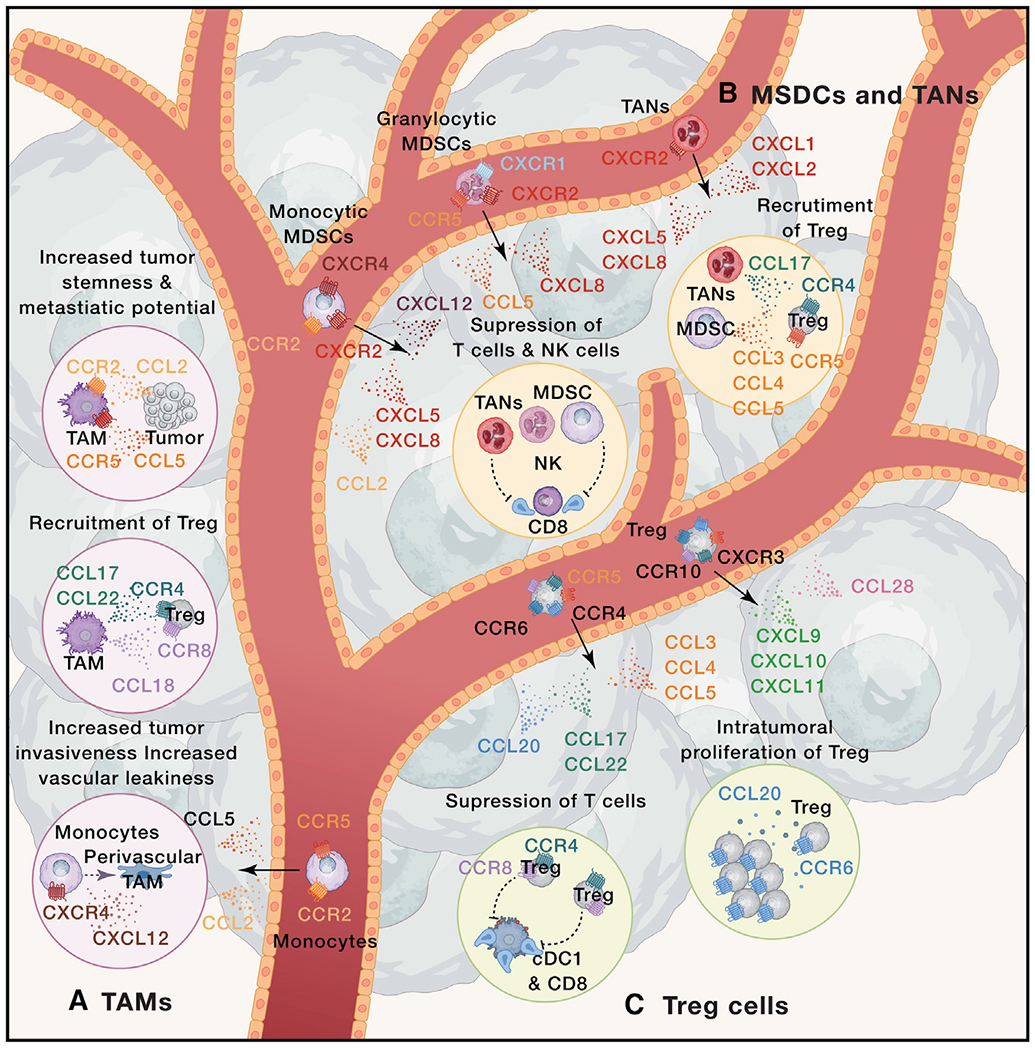
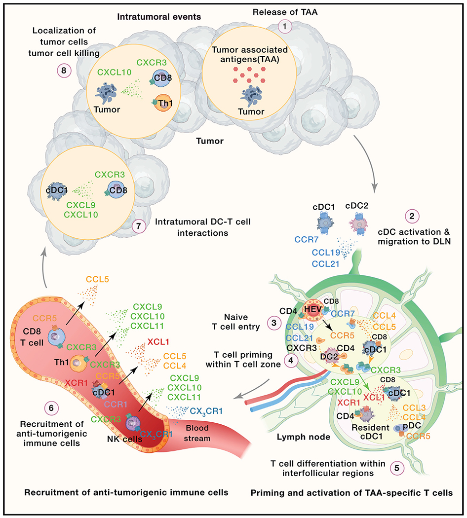
Chemokines are chemotactic cytokines that regulate the migration of immune cells. Chemokines function as cues for the coordinated recruitment of immune cells into and out of tissue and also guide the spatial organization and cellular interactions of immune cells within tissues. Chemokines are critical in directing immune cell migration necessary to mount and then deliver an effective anti-tumor immune response; however, chemokines also participate in the generation and recruitment of immune cells that contribute to a pro-tumorigenic microenvironment. Here, we review the role of the chemokine system in anti-tumor and pro-tumor immune responses and discuss how malignant cells and the tumor microenvironment regulate the overall chemokine landscape to shape the type and outcome of immune responses to cancer and cancer treatment.


Figures

Figure 1.
Figure 1.. Chemokines in pro-tumor immunity
Immune cells, including (A) tumor-associated macrophages (TAMs), (B) myeloid-derived suppressor cells (MDSC), tumor-associated neutrophils (TANs), and (C) regulatory T cells (Treg) contribute to tumor growth. (A) TAMs differentiate from monocytes that are recruited into tumors in response to the CCR5-CCL5 and CCR2-CCL2 chemokine axes. CXCL12 signals through CXCR4 to induce monocyte differentiation towards perivascular macrophages, which contribute to tumor intravasation and vascular leakiness. TAMs contribute to tumor progression through the production of CCL17, CCL22, and CCL18 that attract CCR4+ and CCR8+ Treg cells. CCR2-CCL2 and CCR5-CCL5 chemokine axes regulate cross-talk between TAMs and tumor cells that contributes to tumor stemness and metastatic potential. (B) The CXCR4-CXCL12, CXCR2-CXCL5/CXCL8, and CCR2-CCL2 chemokine axes contribute to the recruitment of monocytic-MDSC, whereas CXCR1-CXCL8, CXCR2-CXCL8, and CCR5-CCL5 axes attract granulocytic-MDSCs. TANs are predominantly recruited through CXCR2 that responds to CXCL1, CXCL2, CXCL5 and CXCL8. Within the TME, MDSCs and TANs suppress T cells and NK cells, as well as contribute to the recruitment of Treg by release of CCL3, CCL4, and CCL5 (MO-MDSCs) or CCL17 (TANs). (C) Treg cells can be recruited by multiple chemokine systems, including CXCR3-CXCL9/CXCL10/CXCL11, CCR10-CCL27, CCR6-CCL20, CCR4-CCL17/CCL22, and CCR5-CCL3/CCL4/CCL5. CCR8+ CCR4+ highly suppressive Treg cells contribute to tumor growth by suppressing T cell responses within TME. Intratumoral Treg cell responses are enhanced by chemokines, such as the CCR6-CCL20 axis, which promotes Treg cell proliferation. Black arrows represent chemokine-mediated cell recruitment. Dotted colored arrows represent cell differentiation and proliferation. Dotted lines represent cell-mediated activation (arrow end) or inhibition (blunt end).
Figure 2.
Figure 2.. Chemokines in anti-tumor immunity
The generation of anti-tumor immunity requires that (1) tumor cells release tumor-associated antigens (TAAs) and neoantigens. (2) TAAs and neoantigens are then taken up and processed by professional antigen-presenting cells (APCs), such as intratumoral conventional type 1 dendritic cells (cDC1s) and cDC2s. In the presence of inflammatory cues, cDCs mature and upregulate CCR7, which promotes their migration into tumor-draining lymph nodes (TDLN) in response to gradients of CCL19 and CCL21. (3) The CCR7-CCL19/CCL21 chemokine axis also controls entry of naive CD8+ and CD4+ T cells into TDLN. (4) Within TDLN, naive TAA-specific CD8+ and CD4+ T cells engage in interactions with cDC1s and cDC2s, respectively. The CCR5-CCL5 chemokine axis facilitates such encounters in the case of naive TAA-specific CD8+ T cells. Activation of CD4+ and CD8+ T cells is accompanied by CXCR3 upregulation, which drives migration of activated T cells into interfollicular regions (IFRs) of the LN. (5) Within IFRs, activated T cells engage in secondary interactions with specific immune cells. For instance, activated CD8+ T cells and migratory cDC1s release chemokines, such as XCL1 and CCL3 and CCL4, to attract LN resident XCR1+ cDC1s and CCR5+ plasmacytoid DCs (pDCs), respectively. TAA-specific CD4+ T cells engage in CXCR3-dependent secondary interactions with DCs that drive their differentiation toward Th1 cells. Moreover, TAA-specific CD4+ T cells and TAA-specific CD8+ T cells interact with the same XCR1+ cDC1s that facilitate the delivery of CD4+ T cell help to primed CD8+ T cells. (6) Various anti-tumorigenic immune ceils, such as NK cells, cDC1 s, Th1 cells, and CD8+ T cells are recruited from the blood into the tumor microenvironment (TME) in response to distinct chemokine gradients. For example, NK cell recruitment is driven by CX3CR1-CX3CL1 and CXCR3-CXCL9/CXCL10/CXCL11. XCR1-XCL1, CCR5-CCL4/CCL5, and CCR1-CCL4 mediate the recruitment of cDC1s. Th1 cells and CD8+ T cells can be recruited through the CXCR3-CXCL9/CXCL10/CXCL11 axis. In addition, CD8+ T cell infiltration can also be triggered by CCR5-CCL5. (7) cDC1s release CXCL9 and CXCL10, contributing to the recruitment of CXCR3+ CD8+ T cells. Moreover, this chemokine system facilitates intratumoral DC-CD8+ T cell interactions, promoting the effector functions of intratumoral Teff cells. (8) Finally, activated CD8+ T cells and Th1 cells localize in the proximity of tumor cells, possibly in response to tumor-derived CXCL10. CD8+ T cells and Th1 cells produce cytokines or directly lyse tumor cells, contributing to tumor eradication. Black arrows represent chemokine-mediated cell recruitment. Gray arrows represent the flow of cells in the tumor immunity cycle. The green arrow represents chemokine-mediated positioning within LNs. Dotted colored arrow represents sequence of cellular events preceding T cell activation and division.
Figure 3.
Figure 3.. Chemokine systems in cancer therapy
Induction of certain chemokines and downregulation of others (solid arrows) in the TME contribute to the efficacy of cancer treatments (dotted arrows), such as radiotherapy, chemotherapy, epigenetic modifiers, and immunotherapy. Moreover, expression of specific chemokine receptors on adoptively transferred T cells plays a critical role in their infiltration into the TME.

References

    1. Acharyya S, Oskarsson T, Vanharanta S, Malladi S, Kim J, Morris PG, Manova-Todorova K, Leversha M, Hogg N, Seshan VE, et al. (2012). A CXCL1 paracrine network links cancer chemoresistance and metastasis. Cell 150, 165–178. - PMC - PubMed
    1. Arwert EN, Harney AS, Entenberg D, Wang Y, Sahai E, Pollard JW, and Condeelis JS (2018). A unidirectional transition from migratory to perivascular macrophage is required for tumor cell intravasation. Cell Rep. 23, 1239–1248. - PMC - PubMed
    1. Ayers M, Lunceford J, Nebozhyn M, Murphy E, Loboda A, Kaufman DR, Albright A, Cheng JD, Kang SP, Shankaran V, et al. (2017). IFN-γ-related mRNA profile predicts clinical response to PD-1 blockade. J. Clin. Invest 127, 2930–2940. - PMC - PubMed
    1. Azizi E, Carr AJ, Plitas G, Cornish AE, Konopacki C, Prabhakaran S, Nainys J, Wu K, Kiseliovas V, Setty M, et al. (2018). Single-cell map of diverse immune phenotypes in the breast tumor microenvironment. Cell 174, 1293–1308.e36. - PMC - PubMed
    1. Balkwill F (2004). Cancer and the chemokine network. Nat. Rev. Cancer 4, 540–550. - PubMed