. 2021 Jan-Jun:296:100650.
doi: 10.1016/j.jbc.2021.100650. Epub 2021 Apr 9.

Interferon regulatory factor 8 regulates expression of acid ceramidase and infection susceptibility in cystic fibrosis

Affiliations

Interferon regulatory factor 8 regulates expression of acid ceramidase and infection susceptibility in cystic fibrosis

Aaron Ions Gardner et al. J Biol Chem. 2021 Jan-Jun.

Abstract

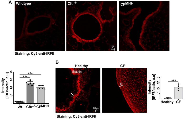
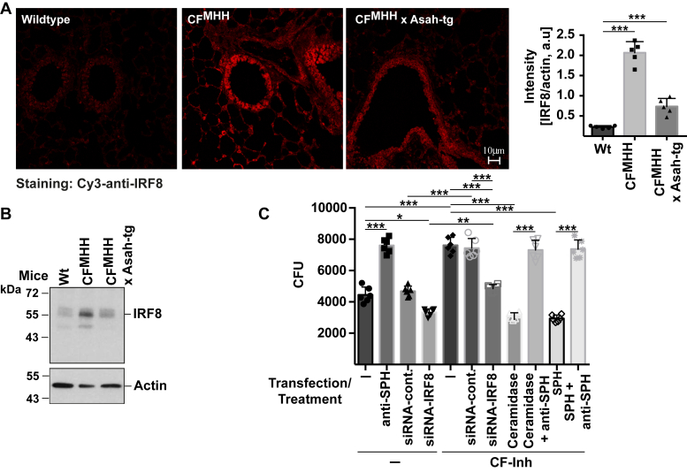
Most patients with cystic fibrosis (CF) suffer from acute and chronic pulmonary infections with bacterial pathogens, which often determine their life quality and expectancy. Previous studies have demonstrated a downregulation of the acid ceramidase in CF epithelial cells resulting in an increase of ceramide and a decrease of sphingosine. Sphingosine kills many bacterial pathogens, and the downregulation of sphingosine seems to determine the infection susceptibility of cystic fibrosis mice and patients. It is presently unknown how deficiency of the cystic fibrosis transmembrane conductance regulator (CFTR) connects to a marked downregulation of the acid ceramidase in human and murine CF epithelial cells. Here, we employed quantitative PCR, western blot analysis, and enzyme activity measurements to study the role of IRF8 for acid ceramidase regulation. We report that genetic deficiency or functional inhibition of CFTR/Cftr results in an upregulation of interferon regulatory factor 8 (IRF8) and a concomitant downregulation of acid ceramidase expression with CF and an increase of ceramide and a reduction of sphingosine levels in tracheal and bronchial epithelial cells from both human individuals or mice. CRISPR/Cas9- or siRNA-mediated downregulation of IRF8 prevented changes of acid ceramidase, ceramide, and sphingosine in CF epithelial cells and restored resistance to Pseudomonas aeruginosa infections, which is one of the most important and common pathogens in lung infection of patients with CF. These studies indicate that CFTR deficiency causes a downregulation of acid ceramidase via upregulation of IRF8, which is a central pathway to control infection susceptibility of CF cells.

Keywords: Pseudomonas aeruginosa; acid ceramidase; ceramide; cystic fibrosis; interferon response factor-8; sphingosine.


Conflict of interest statement

Conflict of interests The authors declare no competing financial interests. M. B., not related to this work: investigator-led research grants from Pfizer and Roche Diagnostics; speaker fees paid to Newcastle University from Novartis, Roche Diagnostics, and TEVA. Travel expenses to educational meetings: Boehringer Ingelheim and Vertex Pharmaceuticals.

Figures

Figure 1
Figure 1
Cftr deficiency induces an upregulation of IRF8—biochemical analysis and a downregulation of acid ceramidase.Left panel, trachea from wild-type (Wt) or Cftr-deficient (Cftr−/−) or Cftr-deficient mice with a residual activity of Cftr (CFMHH) were removed and incubated for 2 h in H/S in the absence or presence of 10 μg/ml P. aeruginosa lipopolysaccharide (LPS). The trachea were then lysed, separated on 7.5% SDS-PAGE, blotted, and analyzed for IRF8 expression. Actin served as loading controls to confirm similar amounts of protein in each lane. Right panel, trachea from 8-week (young) and 24-week (old) wild-type or Cftr−/− mice were removed, immediately lysed, and analyzed as above. All western blots were quantified and normalized to actin expression. Shown are representative blots and the mean ± SD of the quantitative analysis from six independent experiments; ∗∗∗p < 0.001, ANOVA and post hoc t-test.
Figure 2
Figure 2
Cftr/CFTR deficiency induces an upregulation of IRF8—immunofluorescence confocal microscopy.A, trachea from 24-week-old wild-type (Wt) or Cftr−/− or CFMHHmice were removed, fixed in 4% PFA, embedded, sectioned, dewaxed, and immunostained with Cy3-coupled anti-IRF8 antibodies. All figures were made with the same settings of the confocal microscopy. Fluorescence intensity was quantified using Photoshop. IRF8 expression was normalized to actin fluorescence in epithelial cells. Shown are representative immunostainings and the mean ± SD of the quantitative analysis from six each independent samples; ∗∗∗p < 0.001, ANOVA and post hoc t-test. B, sections from explanted lungs from CF patients and from donor lungs were immunostained for IRF8 and analyzed by confocal microscopy. Shown are representative results from six CF lungs and three healthy controls. Fluorescence intensity was quantified using Photoshop and IRF8 expression was normalized to actin fluorescence in epithelial cells. The mean ± SD of the quantitative analysis is given; ∗∗∗p < 0.001, ANOVA and post hoc t-test. E, epithelial cell layer. Please note that the strong signal under the epithelial cell layer in healthy lungs is caused by the autofluorescence of elastin, which is reduced in fibrotic CF lungs.
Figure 3
Figure 3
Cftr/CFTR deficiency induces a downregulation of acid ceramidase, which correlates with increased ceramide and decreased sphingosine levels in tracheal epithelial cells from CF mice.A, acid ceramidase activity in the epithelial cell layer from wild-type (Wt) and CFMHH mice was determined by incubation of the cell surface with [14C]C16ceramide and measuring of its consumption to sphingosine and the fatty acid. Given are the mean ± SD of the acid ceramidase activities from each of the six independent samples; ∗p < 0.05, ∗∗p < 0.01, ∗∗∗p < 0.001, ANOVA and post hoc t-test. B, sections from explanted lungs from CF patients and from donor lungs were immunostained for acid ceramidase and analyzed by confocal microscopy. Shown are representative results from six CF lungs and three healthy controls. Fluorescence intensity was quantified as above and normalized to actin fluorescence in epithelial cells. The mean ± SD of the quantitative analysis is shown; ∗∗∗p < 0.001, ANOVA and post hoc t-test. E, epithelial cell layer. Ceramide (C) and sphingosine (D) in young (8 weeks) and old (24 weeks) wild-type (Wt) and CFMHH were determined in isolated tracheal epithelial cells by kinase assays. Given are the total amounts of ceramide and sphingosine. Shown are the mean ± SD from six independent experiments; ∗∗p < 0.01, ∗∗∗p < 0.001, ANOVA and post hoc t-test.
Figure 4
Figure 4
Pharmacological inhibition of CFTR upregulates IRF8 and downregulates acid ceramidase.A and B, Caco-2 epithelial cells were treated with the pharmacological CFTR Inhibitor-172 (CF-Inh, 1 or 2 μM) for 2 days or left untreated, lysed, and expression of IRF8 was determined by quantitative PCR (A) or western blotting (B). To determine activity (C) and expression (D and E) of the acid ceramidase Caco-2 epithelial cells were treated with the CFTR Inhibitor-172 for 2 days or left untreated, and lysed. C, acid ceramidase activity was measured by consumption of [14C]C16ceramide. Expression of acid ceramidase mRNA (D) and protein (E) (Ac, α and β subunit) was analyzed by quantitative PCR or western blotting. Ceramide (F) and sphingosine (G) levels were analyzed in Caco-2 epithelial cells by kinase assays upon treatment with the CFTR Inhibitor-172 for 2 days. Given are the total amounts of ceramide and sphingosine. Western blots in panel B and E were quantified and normalized to actin expression. Shown are representative blots (B and E) and the mean ± SD of the quantitative studies from each of the six (B, C, E, F and G) or four (A and D) independent experiments; ∗∗∗p < 0.001, ANOVA and post hoc t-test.
Figure 5
Figure 5
CFTR deficiency in differentiated human epithelial cells induces a downregulation of acid ceramidase via an upregulation of IRF8. IRF8-mRNA (A), IRF8 protein levels (B), and acid ceramidase activities (C) were determined by western blotting and enzyme activity assay in cultures of human airway epithelial cells from healthy and CF individuals. To target IRF8 expression, cells were transfected with CRISPR/Cas9 constructs targeting the IRF8 gene (IRF8-CRSPR/Cas9). The density of the IRF8 and acid ceramidase signals in the western blots were quantified using band densitometry. Ceramide levels (D) were determined in untransfected or IRF8-targeted human epithelial cells by a fluorescent activity assay and dot blotting of lipid fractions. The SD is very small in panel A, and we gave the numerical value for easier identification. Shown are mean ± SD of the quantitative studies from each of the four independent experiments; ∗p < 0.05, ∗∗p < 0.01, ∗∗∗p < 0.001, ANOVA and post hoc t-test.
Figure 6
Figure 6
Modification of IRF8 expression regulates acid ceramidase expression. SiRNA-mediated suppression of IRF8 expression results in upregulation of acid ceramidase activity (A), downregulation of ceramide (B), and upregulation of sphingosine (C) in Caco-2 cells expression, while control siRNA has no effect on acid ceramidase, ceramide, and sphingosine (AC). Treatment with the CFTR Inhibitor-172 (CF-Inh, 2 μM) results in an IRF8-dependent downregulation of acid ceramidase activity, an increase of ceramide, and a downregulation of sphingosine, events that are prevented by suppression of IRF8 expression, but not by control (contr.) siRNA. Cells were transiently transfected with siRNA targeting IRF8 or control siRNA by electroporation. Cells were analyzed 48 h after transfection. Cells were left untreated or treated for 2 days with 2 μM CFTR Inhibitor-172. Cells were then lysed and the activity of acid ceramidase and the concentrations of ceramide and sphingosine were determined as above. Expression of IRF8 after transfection of siRNA targeting IRF8 or control siRNA was controlled by western blotting (D). Actin served as loading control. Blots were quantified and normalized to actin. Given are the total amounts of ceramide and sphingosine. Shown are the mean ± SD or representative blots from six independent experiments; ∗p < 0.05, ∗∗∗p < 0.001, ANOVA and post hoc t-test.
Figure 7
Figure 7
Mechanisms of IRF8 upregulation in CF cells, which regulates the cellular defense against P. aeruginosa via sphingosine.A, trachea from wild-type, CF, and and CFxAsah-tg mice were isolated, fixed in 4% paraformaldehyde, embedded in paraplast, sections were performed and stained with Cy3-coupled anti-IRF8 antibodies. The fluorescence signals were quantified and normalized to the fluorescence signal obtained in the epithelial cells for staining with FITC-phalloidin. B, in addition, proteins were extracted and western blots for IRF8 expression were performed. Shown are representative confocal microscopy studies and blots and the mean ± SD of the quantitative analysis from five independent experiments; ∗∗∗p < 0.001, ANOVA and post hoc t-test. C, Caco-2 cells were transiently transfected with siRNA targeting IRF8 or control siRNA and treated with the CFTR Inhibitor-172 (CF-Inh, 2 μM) for 2 days or left untreated. Cells were then infected with P. aeruginosa at a multiplicity of infection (MOI) of 0.1 and the number of the bacteria after 2 h incubation was determined. Surface sphingosine was neutralized by addition of 1 μg/ml anti-sphingosine antibodies added 15 min prior to the infection. Sphingosine on the cell surface was increased by addition of sphingosine (1 μM) or purified neutral ceramidase (1 μg/ml) 15 min prior to infection. Shown are the mean ± SD of the bacterial numbers in the cultures measured as colony forming units (CFU) from six independent experiments; ∗p < 0.05, ∗∗p < 0.01, ∗∗∗p < 0.001, ANOVA and post hoc t-test.

References

    1. Riordan J.R., Rommens J.M., Kerem B., Alon N., Rozmahel R., Grzelczak Z., Zielenski J., Lok S., Plavsic N., Chou J.L., Drumm M.L., Iannuzzi M.C., Collins F.S., Tsui L.C. Identification of the cystic fibrosis gene: Cloning and characterization of complementary DNA. Science. 1989;245:1066–1073. - PubMed
    1. Kerem B., Rommens J.M., Buchanan J.A., Markiewicz D., Cox T.K., Chakravarti A., Buchwald M., Tsui L.C. Identification of the cystic fibrosis gene: Genetic analysis. Science. 1989;245:1073–1080. - PubMed
    1. Elborn J.S. Cystic fibrosis. Lancet. 2016;388:2519–2531. - PubMed
    1. Bhagirath A.Y., Li Y., Somayajula D., Dadashi M., Badr S., Duan K. Cystic fibrosis lung environment and Pseudomonas aeruginosa infection. BMC Pulm. Med. 2016;16:174. - PMC - PubMed
    1. CF Foundation . Cystic Fibrosis Foundation; Bethesda, MD: 2019. Patient Registry Annual Report.

Publication types

MeSH terms