Review
. 2021 Jan-Jun:296:100647.
doi: 10.1016/j.jbc.2021.100647. Epub 2021 Apr 9.

Major advances in targeted protein degradation: PROTACs, LYTACs, and MADTACs

Affiliations
Review

Major advances in targeted protein degradation: PROTACs, LYTACs, and MADTACs

Shanique B Alabi et al. J Biol Chem. 2021 Jan-Jun.

Abstract

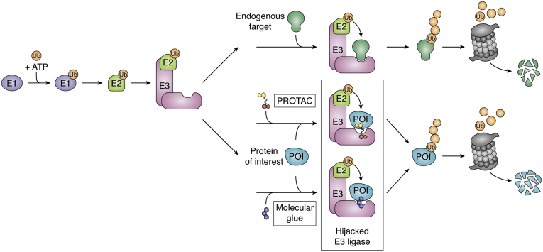
Of late, targeted protein degradation (TPD) has surfaced as a novel and innovative chemical tool and therapeutic modality. By co-opting protein degradation pathways, TPD facilitates complete removal of the protein molecules from within or outside the cell. While the pioneering Proteolysis-Targeting Chimera (PROTAC) technology and molecular glues hijack the ubiquitin-proteasome system, newer modalities co-opt autophagy or the endo-lysosomal pathway. Using this mechanism, TPD is posited to largely expand the druggable space far beyond small-molecule inhibitors. In this review, we discuss the major advances in TPD, highlight our current understanding, and explore outstanding questions in the field.

Keywords: AUTACs; LYTACs; PROTACs; chemical biology; drug action; lysosome; molecular glues; protein degradation; ubiquitination.


Conflict of interest statement

Conflict of interest C. M. C. is founder, shareholder, and consultant to Arvinas, Inc and Halda, LLC, which support research in his laboratory.

Figures

Figure 1
Figure 1
Hijacking the ubiquitin-proteasome system (UPS). In the first step of degradation via the UPS, the E1 activating enzyme forms a thioester bond with ubiquitin in a reaction powered by ATP. Through thioesterification, the ubiquitin molecule is transferred to an E2-conjugating enzyme. Lastly, by simultaneous engagement of both an E2 and the target protein, the RING E3 ligase coordinates the ubiquitination of lysines on the target POI. Polyubiquitinated POIs are then shuttled to the proteasome for degradation. PROTACs and molecular glues hijack the E3 ligase and recruit neosubstrates for ubiquitination and subsequent degradation.
Figure 2
Figure 2
Linkerology and ternary complex formation. The linker length plays an important role in PROTAC-induced degradation. (i) Short linkers that do not allow each target protein to engage its respective ligand will not allow ternary complex formation and thus will fail to induce degradation. (ii) The ideal linker length(s) will allow for strong and stable ternary complexes to form with positive cooperativity. (iii) As linker length increases, due to increased flexibility, the number of possible ternary complex conformers increases. This increased flexibility is tolerated differently by each E3:POI pair. Thus, the“ideal linker” will vary for each neosubstate and E3 pair.
Figure 3
Figure 3
Co-opting the endosomal–lysosome pathway with LYTACs and MoDE-As. LYTACs and MoDE-As tether extracellular or membrane-bound POIs to a recycling receptor, which facilitates internalization and subsequent lysosomal degradation. LYTACs utilize the cation-independent mannose-6-phosphate receptor CI-M6PR, a recycling membrane protein that binds POIs labeled with an MP6n tag. GalNAc-LYTACs and MoDE-As utilize a GalNAC tag to recruit the asialoglycoprotein receptor (ASGPR), a liver-specific lysosomal targeting receptor, for tissue-specific degradation.
Figure 4
Figure 4
Macroautophagy degradation targeting chimeras (MADTACs): AUTACs/ATTECs. AUTACs encoporate FBnG, an autophagy-inducing molecule that mimics S-guanylation. Through an unknown mechanism, AUTACs trigger K63 ubiquitination and lysosomal degradation of POIs. ATTECs are small molecules that link mHTT to LC3, tethering the protein to a phagophore, and similarly induce degradation via autophagy.

References

    1. Balch W.E., Morimoto R.I., Dillin A., Kelly J.W. Adapting proteostasis for disease intervention. Science. 2008;319:916–919. - PubMed
    1. Adhikari B., Bozilovic J., Diebold M., Schwarz J.D., Hofstetter J., Schröder M., Wanior M., Narain A., Vogt M., Dudvarski Stankovic N., Baluapuri A., Schönemann L., Eing L., Bhandare P., Kuster B. PROTAC-mediated degradation reveals a non-catalytic function of AURORA-A kinase. Nat. Chem. Biol. 2020;16:1179–1188. - PMC - PubMed
    1. Powers E.T., Morimoto R.I., Dillin A., Kelly J.W., Balch W.E. Biological and chemical approaches to diseases of proteostasis deficiency. Annu. Rev. Biochem. 2009;78:959–991. - PubMed
    1. Chen B., Retzlaff M., Roos T., Frydman J. Cellular strategies of protein quality control. Cold Spring Harb. Perspect. Biol. 2011;3 - PMC - PubMed
    1. Dissmeyer N., Coux O., Rodriguez M.S., Barrio R. Proteostasis: A European network to break barriers and integrate science on protein homeostasis. Trends Biochem. Sci. 2019;44:383–387. - PubMed

Publication types