. 2021 Jul;9(7):765-778.
doi: 10.1158/2326-6066.CIR-20-0499. Epub 2021 Apr 10.

Exercise Training Improves Tumor Control by Increasing CD8+ T-cell Infiltration via CXCR3 Signaling and Sensitizes Breast Cancer to Immune Checkpoint Blockade

Affiliations

Exercise Training Improves Tumor Control by Increasing CD8+ T-cell Infiltration via CXCR3 Signaling and Sensitizes Breast Cancer to Immune Checkpoint Blockade

Igor L Gomes-Santos et al. Cancer Immunol Res. 2021 Jul.

Abstract

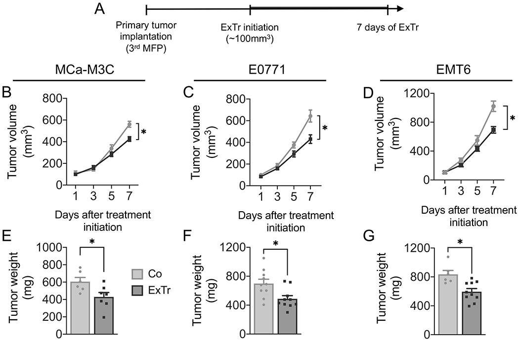
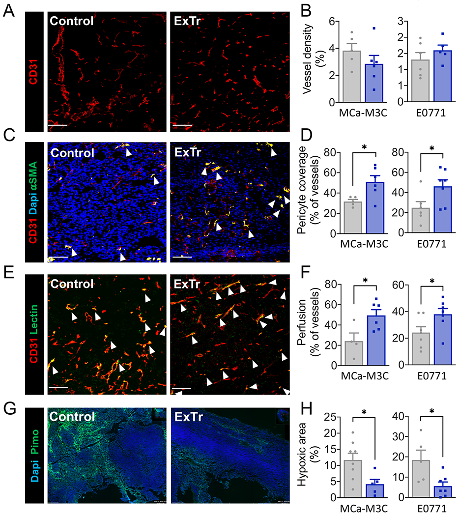
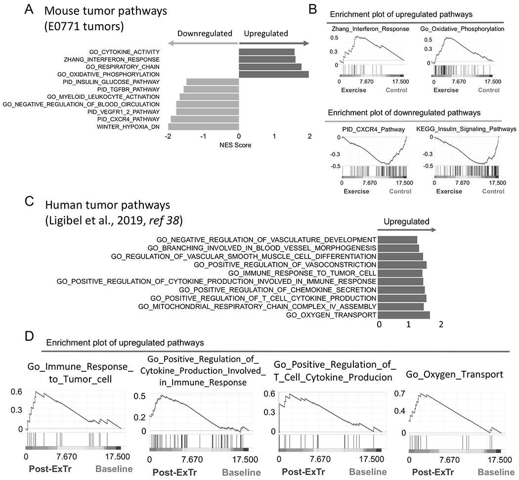
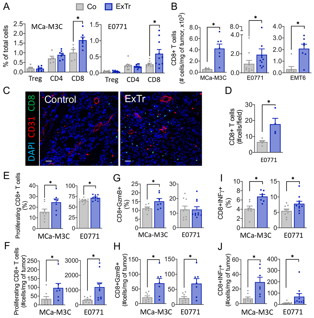
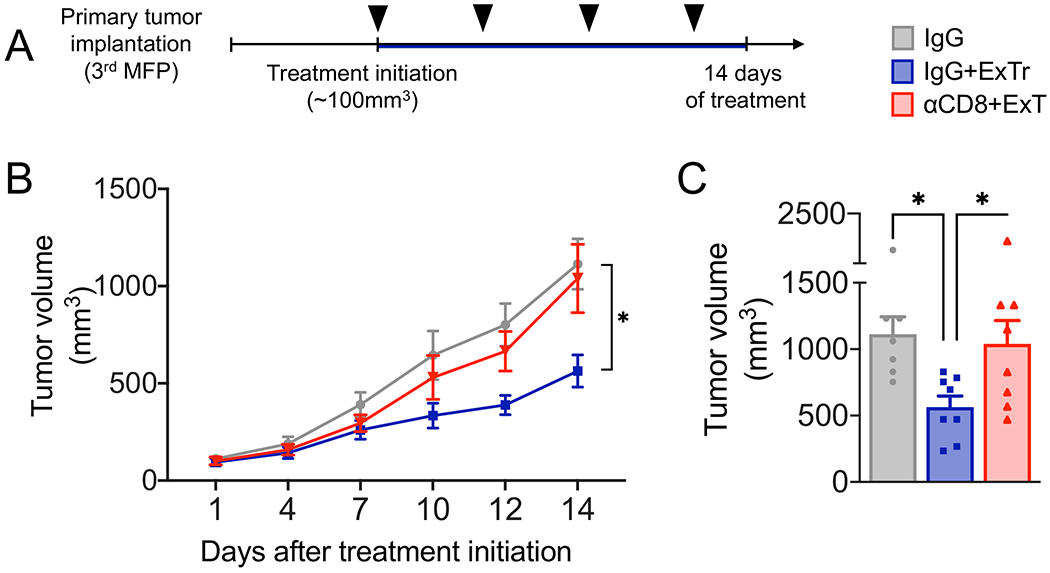
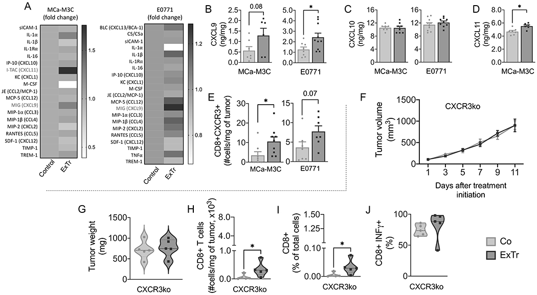
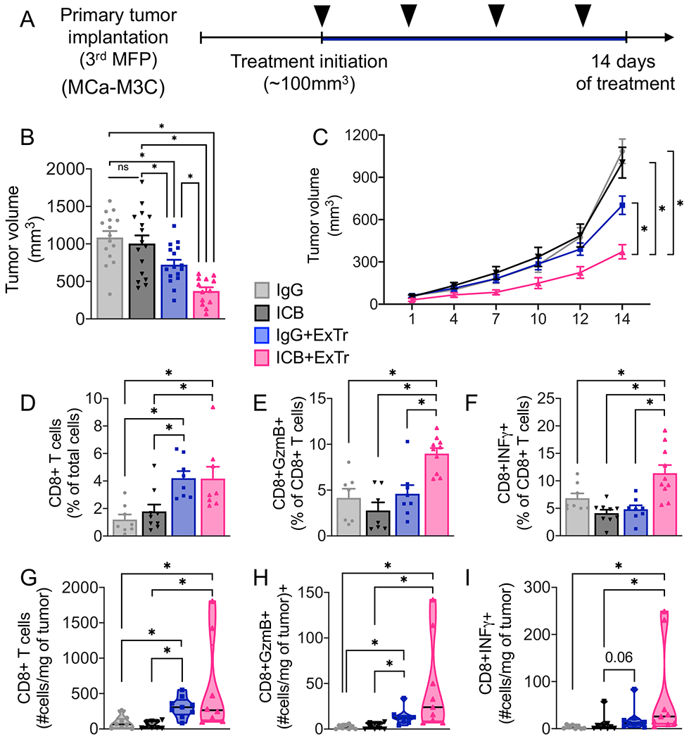
The mechanisms behind the antitumor effects of exercise training (ExTr) are not fully understood. Using mouse models of established breast cancer, we examined here the causal role of CD8+ T cells in the benefit acquired from ExTr in tumor control, as well as the ability of ExTr to improve immunotherapy responses. We implanted E0771, EMT6, MMTV-PyMT, and MCa-M3C breast cancer cells orthotopically in wild-type or Cxcr3-/- female mice and initiated intensity-controlled ExTr sessions when tumors reached approximately 100 mm3 We characterized the tumor microenvironment (TME) using flow cytometry, transcriptome analysis, proteome array, ELISA, and immunohistochemistry. We used antibodies against CD8+ T cells for cell depletion. Treatment with immune checkpoint blockade (ICB) consisted of anti-PD-1 alone or in combination with anti-CTLA-4. ExTr delayed tumor growth and induced vessel normalization, demonstrated by increased pericyte coverage and perfusion and by decreased hypoxia. ExTr boosted CD8+ T-cell infiltration, with enhanced effector function. CD8+ T-cell depletion prevented the antitumor effect of ExTr. The recruitment of CD8+ T cells and the antitumor effects of ExTr were abrogated in Cxcr3-/- mice, supporting the causal role of the CXCL9/CXCL11-CXCR3 pathway. ExTr also sensitized ICB-refractory breast cancers to treatment. Our results indicate that ExTr can normalize the tumor vasculature, reprogram the immune TME, and enhance the antitumor activity mediated by CD8+ T cells via CXCR3, boosting ICB responses. Our findings and mechanistic insights provide a rationale for the clinical translation of ExTr to improve immunotherapy of breast cancer.


Figures

Figure 1.
Figure 1.. Impact of exercise training on tumor progression in established breast tumors
A, Experimental design of the study: MCa-M3C (in FVB), E0771 (in C57BL/6), and EMT6 (in Balb/c) tumors were implanted in the mammary fat pad (MFP) of immunocompetent female mice. When tumors reached ~100 mm3 in size, mice underwent intensity-controlled, moderate-to-vigorous aerobic exercise training (ExTr, 60% of maximal running velocity) or remained sedentary (Control) for 7 consecutive days. B–D, Tumor growth in (B) MCa-M3C (n=6-7), (C) E0771 (n=10), and (D) EMT6 (n=6-10) tumor models in response to 7 days of ExTr. E–G, Tumor weight after 7 days of ExTr the mouse BC models. Statistical comparison by two-way ANOVA (B-D) or unpaired Student’s t test (E-G). Data presented as mean ± SEM.
Figure 2.
Figure 2.. Exercise training normalizes tumor blood vessels and reduces hypoxia.
Immunofluorescence quantification was done in tumor sections to measure vessel normalization and hypoxia. Representative images from E0771 model (A, C, E and G) and corresponding quantitative data (B, D, E and H). A–B, Vessel density, indicated by fractional area (stained for CD31+). C–D, Vessel normalization, characterized by the fraction of vessels with pericyte coverage (CD31+αSMA+). E–F, Perfused vessels, expressed as the fraction of vessels (CD31+) also positive for lectin. G–H, Fractional hypoxic area. Red indicates vessels; Green indicates pericytes (αSMA, C), perfused vessels (lectin, E), or hypoxic area (pimonidazole, G); Blue indicates nucleus (Dapi, C and G). White arrows highlight co-staining. Scale bar, 100 μm. n=4-8 (MCa-M3C) and n=5-7 (E0771). Statistical differences between groups done using unpaired Student’s t test. Data presented as mean ± SEM.
Figure 3.
Figure 3.. Exercise training reprograms the transcription profile in the TME of breast tumors.
A–B, RNA-seq analysis of bulk tumors showing effects of ExTr signaling pathways. (A) Horizontal bars indicate NES (Normalized Enrichment Score) of pathways upregulated (positive NES, in dark color, right) or downregulated (negative NES, in light color, left) by ExTr, indicated by Gene Set Enrichment Analysis (GSEA). (B) Representative GSEA plots showing enhanced molecular signatures. E0771 tumors, collected after 7 days of ExTr. n=5 mice per group, FDR (false discovery rate) q-value<0.12, p<0.05. Comprehensive list of pathways modulated by ExTr are included in the Supplementary section (Supplementary Fig. S5, Supplementary Table S1). C–D, Gene signatures of pathways related to antitumor immunity and vessel normalization in patients in response to ExTr. Original data set from Ligibel et al. (38).
Figure 4.
Figure 4.. Exercise training improves infiltration and effector function of CD8+ T cells in breast tumors.
A, Flow cytometric analysis of tumor infiltrating TCR+CD4+FOXP3+ (Tregs), TCR+CD4+, and TCR+CD8+ T cells, assessed as percentage of total cells in breast tumors. B, Number of CD8 T cells in the three BC models. C, Representative immunofluorescence image [red indicates vessels (CD31+) and green indicates CD8+ T cells] and quantification of tumor immunohistochemistry images of CD8+ T cells per field. Scale bar, 25 μm (n=5 mice per group). D, Absolute number of CD8+ T cells per field. E–J, CD8+ T-cell functions. (E, H) Proliferating (Ki67+) CD8+ T cells. (F, I) Granzyme B+ and (G, J) IFNγ+ CD8+ T cells. For flow cytometry, 2 to 3 independent experiments were performed, and the results are representative of one single experiment (n=6-10 mice per group). Statistical differences comparing groups by unpaired Student’s t test. Data presented as mean ± SEM.
Figure 5.
Figure 5.. CD8+ T cells are required for the antitumor effect of exercise training in established breast tumors.
A, Experimental design of the study. When MCa-M3C tumors reached ~100 mm3, mice underwent daily ExTr (60% of maximal exercise velocity for 14 days) and treatment with anti-CD8β or isotype control IgG (250 μg, i.p., every 4 days; black arrow heads) or kept sedentary with IgG treatment. B, Effects of antibody-mediated CD8+ T-cell depletion (αCD8β) on the antitumor effect of ExTr as measured by tumor growth (B, Two-way ANOVA) and C, final tumor volume (one-way ANOVA), n=7-8 per group. Data presented as mean ± SEM.
Figure 6.
Figure 6.. CXCL9/11-CXCR3 pathway is required for the CD8+ T cell-mediated antitumor effect of exercise training.
A, Heat map of cytokine array on MCa-M3C and E0771 tumors (pooled samples from n=6 mice per group). B–D, ELISA of chemokines (B) CXCL9, (C) CXCL10, and (D) CXCL10 in bulk tumors after 7 days of ExTr (n=6-10 per group, samples run in duplicate). E, Absolute number of CXCR3+CD8+ T cells for both MCa-M3C and E0771 tumor models (flow cytometry). (F-G) We implanted E0771 tumors in Cxcr3−/− mice (C57BL/6 background). When tumors reached ~100 mm3, mice started ExTr (daily sessions at 60% of maximal exercise velocity for 11 days) or were kept sedentary. (F) Tumor growth and (G) final tumor weight in Cxcr3−/− mice. (H) total and (I) relative tumor infiltration of CD8+ T cells in Cxcr3−/− mice. (J) CD8+ T-cell expression of IFNγ in both groups. n=5 mice/group. Statistical differences by comparing groups by unpaired Student’s t test (B-E), two-way ANOVA (F), and Mann-Whitney test (G-J). Data presented as mean ± SEM or median ± interquartile range and distribution (violin plot, G-J).
Figure 7.
Figure 7.. Exercise training sensitizes breast cancer to immune checkpoint blockade.
A, Experimental design of the study. When MCa-M3C tumors reached ~100 mm3, mice underwent ExTr (60% of Vmax ExTr, for 14 days) or kept sedentary and were treated with anti-PD-1 plus anti-CTLA-4 (ICB) or isotype control IgG (i.p., every 4 days; black arrow heads). B, ICB effects on tumor volume. C, The effect of ExTr on MCa-M3C tumor growth with ICB. D–I, TCR+CD8+ T-cell infiltration and function. (D) Relative and (G) absolute numbers of CD8+ T cells. (E) Relative and (H) absolute number of granzyme B+CD8+ T cells. (F) Relative and (I) absolute number of IFNγ+CD8+ T cells with ICB and ExTr. Statistical differences by (B, D-F) one-way or (C) Two-way ANOVA, or (G-I) Kruskal-Wallis test. n=7-16 mice per group, pooled data from 2-3 independent experiments. Data presented as mean ± SEM or median ± interquartile range and distribution (violin plot, G-I).

Similar articles

Cited by

References

    1. Holmes MD, Chen WY, Feskanich D, Kroenke CH, Colditz GA. Physical activity and survival after breast cancer diagnosis. JAMA 2005;293(20):2479–86 doi 10.1001/jama.293.20.2479. - DOI - PubMed
    1. Iyengar NM, Jones LW. Development of Exercise as Interception Therapy for Cancer: A Review. JAMA Oncol 2019. doi 10.1001/jamaoncol.2019.2585. - DOI - PMC - PubMed
    1. Williams PT. Significantly greater reduction in breast cancer mortality from post-diagnosis running than walking. Int J Cancer 2014;135(5):1195–202 doi 10.1002/ijc.28740. - DOI - PubMed
    1. Cannioto RA, Hutson A, Dighe S, McCann W, McCann SE, Zirpoli GR, et al. Physical activity before, during and after chemotherapy for high-risk breast cancer: relationships with survival. J Natl Cancer Inst 2020. doi 10.1093/jnci/djaa046. - DOI - PMC - PubMed
    1. McCullough DJ, Stabley JN, Siemann DW, Behnke BJ. Modulation of blood flow, hypoxia, and vascular function in orthotopic prostate tumors during exercise. J Natl Cancer Inst 2014;106(4):dju036 doi 10.1093/jnci/dju036. - DOI - PMC - PubMed

Publication types

MeSH terms