Review
. 2021;58(4):207-230.
doi: 10.1159/000514211. Epub 2021 Apr 9.

Vascular Patterning as Integrative Readout of Complex Molecular and Physiological Signaling by VESsel GENeration Analysis

Affiliations
Review

Vascular Patterning as Integrative Readout of Complex Molecular and Physiological Signaling by VESsel GENeration Analysis

Mark Lagatuz et al. J Vasc Res. 2021.

Abstract

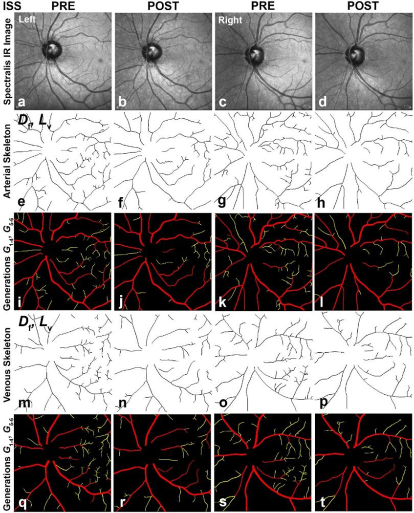
The molecular signaling cascades that regulate angiogenesis and microvascular remodeling are fundamental to normal development, healthy physiology, and pathologies such as inflammation and cancer. Yet quantifying such complex, fractally branching vascular patterns remains difficult. We review application of NASA's globally available, freely downloadable VESsel GENeration (VESGEN) Analysis software to numerous examples of 2D vascular trees, networks, and tree-network composites. Upon input of a binary vascular image, automated output includes informative vascular maps and quantification of parameters such as tortuosity, fractal dimension, vessel diameter, area, length, number, and branch point. Previous research has demonstrated that cytokines and therapeutics such as vascular endothelial growth factor, basic fibroblast growth factor (fibroblast growth factor-2), transforming growth factor-beta-1, and steroid triamcinolone acetonide specify unique "fingerprint" or "biomarker" vascular patterns that integrate dominant signaling with physiological response. In vivo experimental examples described here include vascular response to keratinocyte growth factor, a novel vessel tortuosity factor; angiogenic inhibition in humanized tumor xenografts by the anti-angiogenesis drug leronlimab; intestinal vascular inflammation with probiotic protection by Saccharomyces boulardii, and a workflow programming of vascular architecture for 3D bioprinting of regenerative tissues from 2D images. Microvascular remodeling in the human retina is described for astronaut risks in microgravity, vessel tortuosity in diabetic retinopathy, and venous occlusive disease.

Keywords: 3D bioprinting; Angiogenesis; Central retinal vein occlusion; Diabetic retinopathy; Keratinocyte growth factor; Leronlimab; Microvascular; Saccharomyces boulardii; Spaceflight-Associated Neuro-ocular Syndrome.


Conflict of interest statement

Conflict of Interest Statement

The authors have no conflicting or competing interests to disclose.

Figures

Fig. 1.
Fig. 1.. Mapping and quantification by VESGEN of arterial and venous trees.
The vascular tree analysis is illustrated by an image of the left retina of an astronaut crew member acquired after a 6-month mission to the ISS. Binary arterial and venous trees extracted from the grayscale image (a–c) served as sole inputs to the software for the automated generation of arterial and venous maps (d–i). The software first calculates a skeleton (centerline) of the vascular branching that supports calculation of the fractal dimension (Df) and overall vessel length density (Lv). Together the vascular binary image and skeleton then generate additional mappings such as the distance map and branching generations for calculation of site-specific information on differences in vessel parameters that include small and large vessel diameter, density, and tortuosity. As shown by the legend, colors within the distance map correspond to pixel distance to the edge of the vessel. Arterial and venous Df were 1.354 and 1.329 and Lv, 12.1×10⁻⁴ and 10.7×10⁻⁴ μm/μm2. Average diameter ± SD of arteries and veins per branching generation ranged from 35.5 ± 1.8 and 41.1 ± 3.9 μm for G1 (largest generation) to 13.2 ± 2.1 and 13.0 ± 11.6 μm for G5 (smallest). Arterial and venous branching generations (F, I) can be grouped by a user option into groups such as large and small vessels (Fig. 2). Scalebar, 200 μm. VESGEN, VESsel GENeration; ISS, International Space Station; IR, infrared.
Fig. 2.
Fig. 2.. Vascular decrease in the retinas of an astronaut after 6 months on the ISS.
Arterial and venous density decreased in the left and right post-flight retinas of an astronaut crew member. Binary arterial and venous trees extracted from grayscale images of the left and right retinas (a–d) were analyzed by VESGEN as vascular skeletons (e–h, m–p) and generational branching grouped into large (Gv1–4, red) and small (Gv5–6, yellow) generations (i–l, q–t). Decreases in vascular parameters were calculated from skeletonized and grouped maps (Table 1). For visual comparison, all images were aligned with optic nerve to left (i.e., images of right retina rotated 180°). Scalebars, 200 μm.
Fig. 3
Fig. 3. Remodeling of arterial and venous trees with progression of nonproliferative diabetic retinopathy (NPDR).
Clinical images of the human retina with mild (a, d) and moderate (G) stages of NPDR by the increasing presence of established clinical disease markers such as microaneurysms and hemorrhagic leakage. One image (d) is representative of a subset of retinas within the study (approximately 13%) displaying the additional phenotype of unusually tortuous vessels (Table 2). Arterial (b, e, h) and venous (c, f, i) maps illustrate branching generations from G1 to G7 (legend). The generational maps were grouped into large (G1–4) and small (G≥5) generations to further quantify site-specific changes within the complex branching trees using the VESGEN generation grouping feature (Table 2). Scalebars, 200 μm.
Fig. 4
Fig. 4. Epithelial activator KGF induces vessel tortuosity in vivo.
The quail CAM, avian analog of the placenta, was treated (a) with and (d) without KGF at 10 μg CAM for 48 hr. Images generated by VESGEN of the arterial end points include distance maps (b, e) where legend indicates pixel distance to vessel edge and branching generations with legend (G1G7; c, f) that support quantification of specific generational changes exerted by KGF (Table 3). As a novel regulator of vessel tortuosity, KGF may have increased the activity of matrix metalloproteases in the chorionic epithelium [50], thereby decreasing tissue resistance to sinusoidal vascular patterning by the pulsatile blood flow. Scalebar, 500 μm (d).
Fig. 5.
Fig. 5.. Inhibition of tumor angiogenesis in a humanized mouse model.
Immuno-incompetent mice (NSG) were humanized by inoculation with normal human bone marrow derived leukocytes. Then SW480 human colon carcinoma cells were injected intradermally in the mouse flanks. Mice were treated either with normal human IgG (left columns) or leronlimab (right columns) at a dose of 2 mg/kg i.p. twice weekly. On day 10 the mice were euthanized, and the tumor inoculation site was photographed and subjected to VESGEN analysis.
Fig. 6.
Fig. 6.. Network analysis of pathological angiogenesis with probiotic protection during intestinal inflammation.
(first row) In confocal fluorescent images, the luminal capillary network of the normal mouse colon appears as a regular lattice structure (left column) that is disrupted by inflammation following administration of DSS (middle column). Treatment with S. boulardii, a yeast probiotic (right column), moderated the inflammatory angiogenic response. (second row) Binary vascular patterns extracted from confocal images were mapped and quantified as described previously with the Vascular Network option [32], in which the entire vascular pattern is analyzed for vessel parameters and fractional vascular/avascular areas (Table 4). However, only regions containing complete AVS (black) are quantified for other network parameters. Scalebar, 300 μm.
Fig. 7.
Fig. 7.. Tree-Network Analysis of microvascular changes in the human retina from CRVO.
Vessels in the parafoveal region of the human retina were imaged by AOSLO-FA [33] and analyzed by the VESGEN Tree-Network Composite option. Compared to the control retina of a healthy young adult, vessel density within the CRVO retina of this middle-aged subject was reduced and irregular (Table 5). Vascular damage was accompanied by prominent microaneurysms, considerable fluorescein leakage from the vessels and apparent vascular dropout in the central avascular foveal region. Vessel changes in the fellow retina of the CRVO subject were intermediate between that of the control and CRVO. Scalebar, 300 μm.
Fig. 8.
Fig. 8.. Branch-specific ‘direct write’ 3D printing of a vascular region from a rat retina.
(a) Original fluorescent image. (b) Same image after inversion, segmentation, and smoothing (scalebar, 100 μm). (c) Determination of branching generations by VESGEN (legend). By the analysis, not all branching generations were present, such as G2 and G3. (d) Same image after grouping of generation classes into large (G1–3, red), medium (G4, green) and small (G≥5, blue). (e) CAD conversion of the same field into a .stl printable file. (f) Direct-write printing of the vascular pattern with a bioink surrogate.
Fig. 9.
Fig. 9.
Fractal patterning of self-similar branching in the aqueous transport of river systems and conduction of electrical charge in lightning Photographs of (left) arterial river branching in the Sundarbans coast at the Mouths of the Ganges by the Bay of Bengal [82] and (right) lightning at night [83] illustrate the contrasting physics of self-similar fractal branching in the transport of aqueous fluids such as rivers and biological vascular systems, compared to the conduction of electricity in lightning and neurons of nervous systems. Self-similarity is a fractal pattern whereby the characteristic pattern (in these examples, bifurcational branching) is repeated at increasingly smaller length scales. Unlike many mathematical fractals, fractals in biology and nature are irregular.

References

    1. Folkman J (2007) Angiogenesis: an organizing principle for drug discovery? Nat Rev Drug Discov 6 (4):273–286 - PubMed
    1. Potente M, Gerhardt H, Carmeliet P (2011) Basic and therapeutic aspects of angiogenesis. Cell 146 (6):873–887. doi:10.1016/j.cell.2011.08.039 - DOI - PubMed
    1. De Spiegelaere W, Casteleyn C, Van den Broeck W, Plendl J, Bahramsoltani M, Simoens P, Djonov V, Cornillie P (2012) Intussusceptive angiogenesis: a biologically relevant form of angiogenesis. J Vasc Res 49 (5):390–404. doi:10.1159/000338278 - DOI - PubMed
    1. Wilhelm K, Happel K, Eelen G, Schoors S, Oellerich MF, Lim R, Zimmermann B, Aspalter IM, Franco CA, Boettger T, Braun T, Fruttiger M, Rajewsky K, Keller C, Bruning JC, Gerhardt H, Carmeliet P, Potente M (2016) FOXO1 couples metabolic activity and growth state in the vascular endothelium. Nature 529 (7585):216–220. doi:10.1038/nature16498 - DOI - PMC - PubMed
    1. Apte RS, Chen DS, Ferrara N (2019) VEGF in Signaling and Disease: Beyond Discovery and Development. Cell 176 (6):1248–1264. doi:10.1016/j.cell.2019.01.021 - DOI - PMC - PubMed

Publication types

MeSH terms

Substances