Review
. 2021 Aug;39(8):733-740.
doi: 10.1007/s11604-021-01115-7. Epub 2021 Apr 11.

Congenital anomalies causing hemato/hydrocolpos: imaging findings, treatments, and outcomes

Affiliations
Review

Congenital anomalies causing hemato/hydrocolpos: imaging findings, treatments, and outcomes

Keizo Tanitame et al. Jpn J Radiol. 2021 Aug.

Abstract

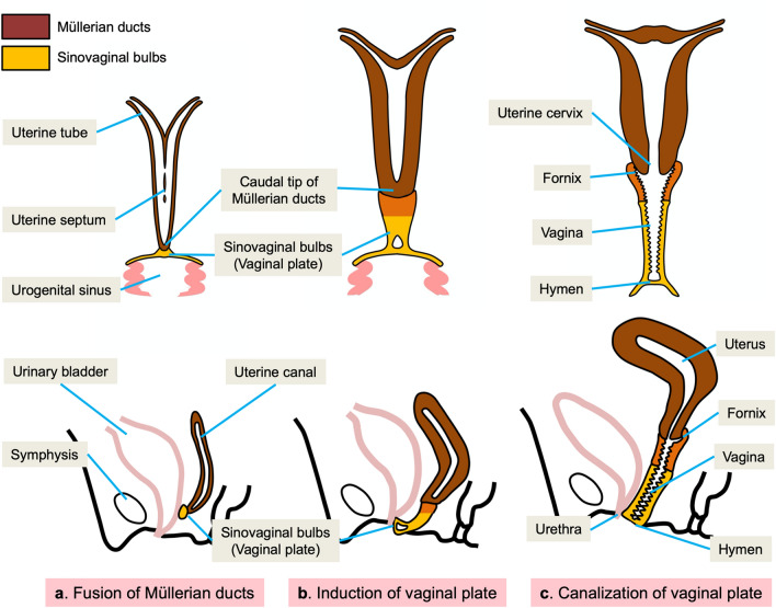
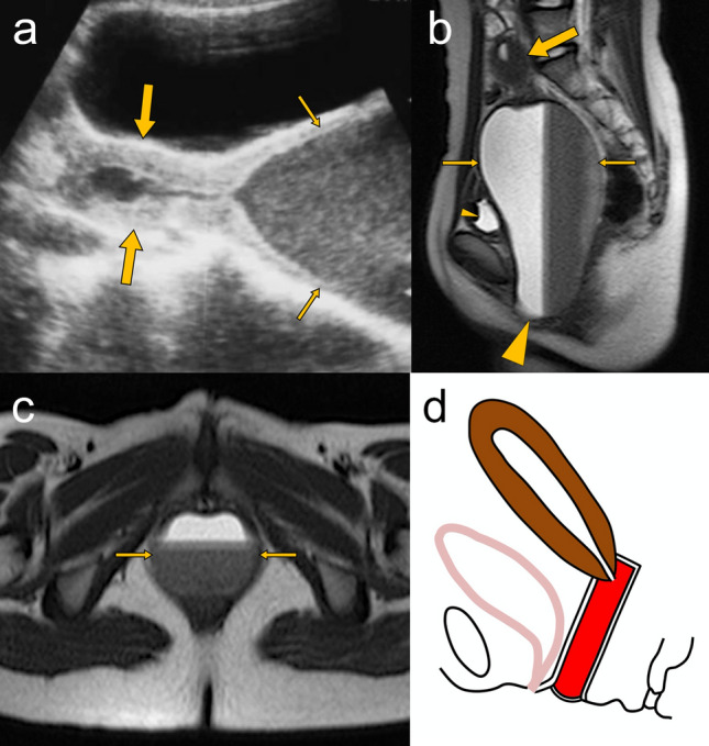
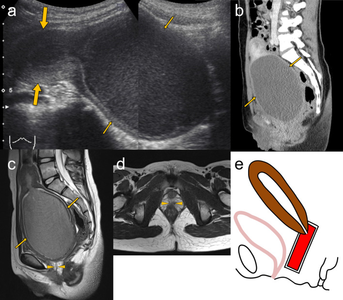
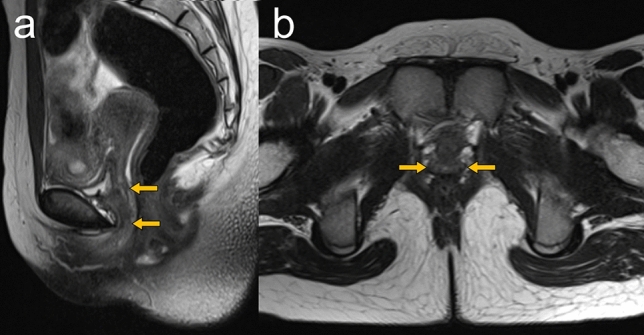
Hemato/hydrocolpos due to congenital urogenital anomalies are rare conditions discovered in neonatal, infant, and adolescent girls. Diagnosis is often missed or delayed owing to its rare incidence and nonspecific symptoms. If early correct diagnosis and treatment cannot be performed, late complications such as tubal adhesion, pelvic endometriosis, and infertility may develop. Congenital urogenital anomalies causing hemato/hydrocolpos are mainly of four types: imperforate hymen, distal vaginal agenesis, transverse vaginal septum, and obstructed hemivagina and ipsilateral renal anomaly, and clinicians should have adequate knowledge about these anomalies. This article aimed to review the diagnosis and treatment of these urogenital anomalies by describing embryology, clinical presentation, imaging findings, surgical management, and postoperative outcomes.

Keywords: Distal vaginal agenesis; Hematocolpos; Imperforate hymen; OHVIRA; Transverse vaginal septum.


Conflict of interest statement

The authors declare that there is no conflict of interest.

Figures

Fig. 1
Fig. 1
Schematic illustrations show coronal views of congenital urogenital anomalies causing hematocolpos. The accumulated blood in the vagina is colored in red. a Imperforate hymen. b Distal vaginal agenesis. c Complete transverse vaginal septum. d Obstructed hemivagina and ipsilateral renal anomaly (OHVIRA)
Fig. 2
Fig. 2
Development of the vagina. a After the caudal tip of the fused Müllerian ducts reaches the urogenital sinus, a sinovaginal bulb grows out of the sinus. b The sinovaginal bulb proliferates and forms a solid vaginal plate. Proliferation continues at the cranial end of the plate. c By the 5th month, the vaginal plate is entirely canalized and forms the vagina
Fig. 3
Fig. 3
A 13-year-old girl with imperforate hymen. a Abdominal ultrasonography shows a normal-shaped uterus (large arrows) and a distended vagina (small arrows) containing echogenic fluid. b Sagittal T2-weighted MRI image shows the distended vagina (small arrows) measuring 14.5 × 7.5 cm in size with a fluid–fluid level (“hematocrit effect”), which compresses the bladder (small arrowhead). The bulging imperforate hymen (large arrowhead) protrudes between the labia. The uterus (large arrow) is normally visualized. c Axial T2-weighted MRI image shows the distended vagina (small arrows) containing blood products between the urethra and anal canal. d Schematic illustration shows a sagittal view of hematocolpos due to imperforate hymen. Intravaginal blood is colored in red
Fig. 4
Fig. 4
An 11-year-old girl with lower vaginal agenesis. a Abdominal ultrasonography shows a normal-shaped uterus (large arrows) and a grossly distended upper vaginal part (small arrows) with moving internal echoes. b Sagittal reformatted contrast-enhanced CT image shows the distended upper part of the vagina (small arrows) containing slightly hyperdense fluid. c, d Sagittal (c) and axial (d) T2-weighted MRI images show the distended upper vagina (small arrows) containing blood products and absence of the lower vagina with replacement by small fibrous tissue (arrowheads). MRI is better than CT in demonstrating distal vaginal agenesis. e Schematic illustration shows a sagittal view of hematocolpos due to distal vaginal agenesis. The accumulated blood in the upper part of the vagina is colored in red
Fig. 5
Fig. 5
An 11-year-old girl after vaginoplasty for distal vaginal agenesis. Sagittal (a) and axial (b) T2-weighted MRI images show a neovagina (arrows). No postoperative complications, such as hydrometra due to vaginal stenosis or vaginal shortening, are observed
Fig. 6
Fig. 6
Instilling of jelly through the vaginal introitus for evaluating transverse vaginal septum. a Schematic illustration shows a sagittal view of hematometrocolpos due to transverse vaginal septum. There is a difficulty to distinguish the vaginal septum from the collapsed lower vagina. b After instilling of jelly through the vaginal introitus, hematometrocolpos (colored in red), the vaginal septum (arrow), and the lower vaginal segment containing jelly (colored in blue) are clearly shown. Intravaginal infusion of jelly has been reported to provide information on the level and thickness of the vaginal septum
Fig. 7
Fig. 7
A 30-year-old pregnant woman at 18 weeks of gestation having right obstructed hemivagina and ipsilateral renal anomaly (OHVIRA) with pyometrocolpos. a Transvaginal ultrasonography shows a distended right hemivagina containing hypoechoic fluid (arrows). bf Axial (b, c) and coronal (d, e) T2-weighted MRI images show duplication of the uterine and vaginal canals. The distended right hemivagina (large arrows), right uterine horn (small arrows), vaginal retention cysts (arrowheads), and a fetus at 17 weeks in the left uterine horn are identified. Coronal T2-weighted MRI image (f) also demonstrates the absence of the right kidney (oval circle). g Schematic illustration shows a coronal view of the right-sided OHVIRA with pregnancy in the left uterus

References

    1. Berkowitz CD, Elvik SL, Logan M. A simulated "acquired" imperforate hymen following the genital trauma of sexual abuse. Clin Pediatr (Phila) 1987;26:307–309. doi: 10.1177/000992288702600609. - DOI - PubMed
    1. Winderl LM, Silverman RK. Prenatal diagnosis of congenital imperforate hymen. Obstet Gynecol. 1995;85:857–860. doi: 10.1016/0029-7844(94)00405-3. - DOI - PubMed
    1. Bajaj SK, Misra R, Thukral BB, Gupta R. OHVIRA: uterus didelphys, blind hemivagina and ipsilateral renal agenesis: advantage MRI. J Hum Reprod Sci. 2012;5:67–70. doi: 10.4103/0974-1208.97811. - DOI - PMC - PubMed
    1. Chircop R. A case of retention of urine and haematocolpometra. Eur J Emerg Med. 2003;10:244–245. doi: 10.1097/00063110-200309000-00019. - DOI - PubMed
    1. Fischer JW, Kwan CW. Emergency point-of-care ultrasound diagnosis of hematocolpometra and imperforate hymen in the pediatric emergency department. Pediatr Emerg Care. 2014;30:128–130. doi: 10.1097/PEC.0000000000000080. - DOI - PubMed

LinkOut - more resources