Review
. 2021;97(4):161-196.
doi: 10.2183/pjab.97.010.

Lipid hydroperoxides in nutrition, health, and diseases

Affiliations
Review

Lipid hydroperoxides in nutrition, health, and diseases

Teruo Miyazawa. Proc Jpn Acad Ser B Phys Biol Sci. 2021.

Abstract

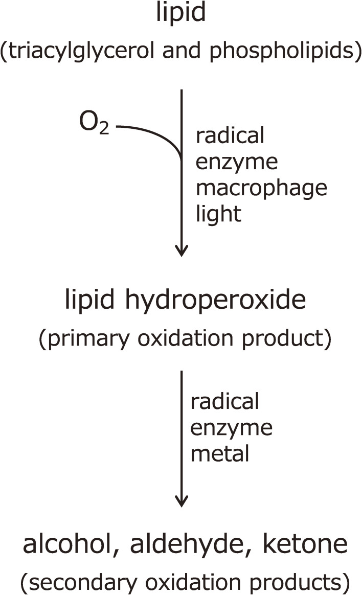
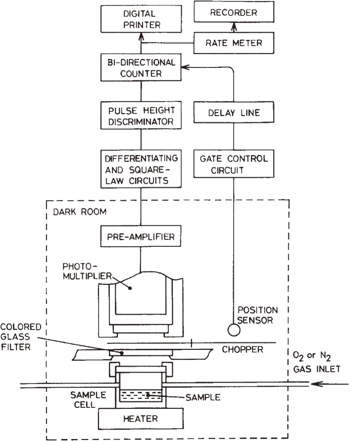
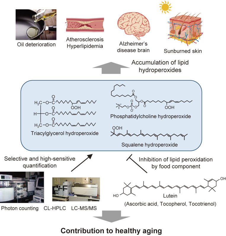
Research on lipid peroxidation in food degradation, oil and fat nutrition, and age-related diseases has gained significant international attention for the view of improvement of societal health and longevity. In order to promote basic studies on these topics, a chemiluminescence detection-high performance liquid chromatography instrument using a high-sensitivity single photon counter as a detector was developed. This instrument enabled us to selectively detect and quantify lipid hydroperoxides, a primary product of lipid peroxidation reactions, as hydroperoxide groups at the lipid class level. Furthermore, an analytical method using liquid chromatography-tandem mass spectrometry has been established to discriminate the position and stereoisomerization of hydroperoxide groups in lipid hydroperoxides. Using these two methods, the reaction mechanisms of lipid peroxidation in food and in the body have been confirmed.

Keywords: Alzheimer’s disease; atherosclerosis; chemiluminescence; immunocompetent cells; lipid hydroperoxides; rancid cooking oil.


Figures

Figure 1.
Figure 1.
Lipid peroxidation reaction and its products.
Figure 2.
Figure 2.
Schematic diagram of the ultra-high sensitivity photon counting system specially designed for biomedical and clinical applications. Adapted with permission from Ref. . Copyright 1982 Elsevier.
Figure 3.
Figure 3.
Chemiluminescence emission spectra observed between phosphatidylcholine hydroperoxide (PCOOH) and cytochrome c. The spectra were recorded using a filter spectral analyzer by mixing 1 M PCOOH with cytochrome c (1 µg/mL) in the presence (broken line) and absence (solid line) of 5 mM β-carotene (a singlet oxygen quencher). Adapted with permission from Ref. . Copyright 1990 John Wiley and Sons.
Figure 4.
Figure 4.
Schematic diagram of chemiluminescence detection-high performance liquid chromatography (CL-HPLC) for the lipid hydroperoxides assay. A, mobile phase; P, pump; I, sample injection valve; S, sample; C, column in a column oven; U, UV detector; J, mixing joint; B, chemiluminescence reagent consisting of cytochrome c and luminol in borate buffer; Q, spiral Teflon cell; PM, photomultiplier; CL, chemiluminescence detector; R, multiple recorder and integrator; W, waste. Adapted with permission from Ref. . Copyright 1990 John Wiley and Sons.
Figure 5.
Figure 5.
Changes in the phosphatidylcholine hydroperoxide (PCOOH) content per 106 cells as a function of the population doubling level (PDL) of human fetal diploid. Values are the mean of three experiments, with SD. The figure in parentheses is the value relative to young cells (20th PDL). Adapted with permission from Ref. . Copyright 1993 John Wiley and Sons.
Figure 6.
Figure 6.
Chemiluminescence detection-high performance liquid chromatography (CL-HPLC) of trioleoylglycerol (peroxide value 0.16 meq/kg). The HPLC column was a Finepak SIL C18-5 (5 µm, 250 × 4.6 mm) connected with a Finepak SIL C18 T-P pre-column (5 µm, 50 × 4.6 mm). The mobile phase was methanol, and the flow rate was 1.1 mL/min. The chemiluminescence reagent was composed of cytochrome c and luminol. Mono-OOH, mono-hydroperoxides (retention time 21.0 min); Bis-OOH, bis-hydroperoxides (retention time 10.0 min); UV, ultraviolet. Adapted with permission from Ref. . Copyright 1995 John Wiley and Sons.
Figure 7.
Figure 7.
Chemiluminescence detection-high performance liquid chromatography (CL-HPLC) chromatograms of soybean oil (A, peroxide value [PV] 6 meq/kg; B, PV 110 meq/kg; C, PV 780 meq/kg) autoxidized at 25 ℃. Indicators, ×1, ×1 : 20, ×1 : 200, are the relative dilution ratios of oxidized oil. LLL, OLL, PLL, OOL, and POL depicted the fatty acid combination in the molecular species of triacylglycerol, with abbreviations representing palmitic (P), oleic (O), and linoleic (L) acids. Adapted with permission from Ref. . Copyright 1995 John Wiley and Sons.
Figure 8.
Figure 8.
Chemical reaction of 1-palmitoyl-2-oleoyl-sn-glycero-3-phosphocholine (POPC) with ozone. PC-ald, 1-palmitoyl-2-(9-oxononanoyl)-sn-glycero-3-phosphocholine; PC-acid, 1-palmitoyl-2-(9-carboxynanoyl)-sn-glycero-3-phosphocholine. Adapted with permission from Ref. . Copyright 2002 John Wiley and Sons.
Figure 9.
Figure 9.
Proposed pathway for the formation of (A) phosphatidylcholine ethoxyhydroperoxide (PC-EHP) and (B) PC-CE during the ozonation of 1-palmitoyl-2-oleoyl-sn-glycero-3-phosphocholine (POPC) in ethanol. For abbreviations, see Fig. 8. Adapted with permission from Ref. . Copyright 2002 John Wiley and Sons.
Figure 10.
Figure 10.
Formation of 7α-ethoxy-5-OOH (7α-ethoxy-3β-hydroxy-5α-B-homo-6-oxacholestene-5-hydroperoxide) during cholesterol ozonation in the presence of ethanol. Adapted with permission from Ref. . Copyright 2004 John Wiley and Sons.
Figure 11.
Figure 11.
Chemical structures of squalene (SQ) and six monohydroperoxide (SQOOH) isomers formed by sunlight exposure. Adapted from Ref. .
Figure 12.
Figure 12.
Predicted pathways of the singlet oxygen oxidation of squalene (SQ) (A) and free radical oxidation of SQ (B). Adapted from Ref. .
Figure 13.
Figure 13.
Correlation between plasma phosphatidylcholine hydroperoxide (PCOOH) and age. ●, control subjects (r = 0.392; P < 0.01); ○, patient with hyperlipidemia (r = 0.298; P < 0.01). Adapted with permission from Ref. . Copyright 2000 Oxford University Press.
Figure 14.
Figure 14.
Involvement of Rac activation in phosphatidylcholine hydroperoxide (PCOOH)-induced THP-1 monocytic cell adhesion to intracellular adhesion molecule-1 (ICAM-1) in atherogenicity.
Figure 15.
Figure 15.
Plasma epigallocatechin gallate (EGCg) (A) and phosphatidylcholine hydroperoxide (PCOOH) (B) before tea catechin administration and 60 min after administration in humans. Values represent the mean ± SD of 18 subjects. Green tea extract (equivalent to 254 mg catechin) was administrated after 12 h of fasting. Adapted with permission from Ref. . Copyright 1999 American Chemical Society.
Figure 16.
Figure 16.
Lipid oxidation mechanism and isomeric structure. Lipid oxidation mechanism (radical, singlet oxygen, or enzymatic) can be estimated by analyzing the structures around the hydroperoxyl group.
Figure 17.
Figure 17.
A: protocols investigated for the purification of lipid hydroperoxide (LOOH) using 2-methoxypropene (MxP). B: chemical structures of lipids [1-palmitoyl-2-linoleoyl-sn-glycero-3-phosphocholine (PLPC), 1-palmitoyl-2-linoleoyl-sn-glycero-3-phosphoethanolamine (PLPE), 1-palmitoyl-2-linoleoyl-sn-glycero-3-phosphoserine (PLPS), cholesteryl linoleate (ChL), trilinoleoylglycerol (LLL), linoleic acid (LA), and methyl linoleate (LAMe)]. Adapted with permission from Ref. . Copyright 2008 American Society for Biochemistry and Molecular Biology.
Figure 18.
Figure 18.
Presumed mechanism of the reaction between 2-methoxypropene (MxP) and hydroperoxide. (A): addition of MxP by nucleophilic addition of hydroperoxide to 2-methoxypropene. (B): elimination of MxP and regeneration of hydroperoxide. Adapted with permission from Ref. . Copyright 2008 American Society for Biochemistry and Molecular Biology.
Figure 19.
Figure 19.
Chemical structures of phosphatidylcholine (PC) and phosphatidylcholine hydroperoxide (PCOOH). 13(S)-9Z,11E-HPODE PC is produced by enzymatic oxidation of PC (1-palmitoyl-2-linoleoyl-sn-glycero-3-phosphocholine (16 : 0/18 : 2 PC)). 13(RS)-9Z,11E-HPODE PC and 13(RS)-9E,11E-HPODE PC are formed by auto-oxidation of PC (A). The preparation scheme for the enzymatic oxidation product and the auto-oxidation products from LA is shown in (B). Adapted with permission from Ref. . Copyright 2016 Springer Nature.
Figure 20.
Figure 20.
Diastereomer analysis of phosphatidylcholine hydroperoxide (PCOOH) bearing 13-9Z,11E-HPODE. PCOOH, prepared via lipoxygenase (LOX)-catalyzed oxidation or auto-oxidation, was subjected to stereoselective liquid chromatography ultraviolet mass spectrometry (LC-UV-MS) to evaluate the R/S configuration of the hydroperoxy group in the PCOOH molecule. PCOOH was detected by UV (234 nm; A) and structure-selective selected reaction monitoring (m/z 812.5/724.4; B). In addition, we prepared PCOOH by directly treating phosphatidylcholine (1-palmitoyl-2-linoleoyl-sn-glycero-3-phosphocholine, 16 : 0/18 : 2 PC) with LOX. The resultant PCOOH was analyzed by stereoselective LC-UV-MS. Adapted with permission from Ref. . Copyright 2015 Elsevier.
Figure 21.
Figure 21.
Diastereomer and/or cis-trans separation of the mixture of 13(RS)-9Z,11E-HPODE PC and 13(RS)-9E,11E-HPODE PC (chromatogram A), 13(S)-9Z,11E-HPODE PC (chromatogram B), 13(RS)-9Z,11E-HPODE PC (chromatogram C), and 13(RS)-9E,11E-HPODE PC (chromatogram D) using a combination of CHIRALPAK OP (+) and IB-3 columns. PCOOH isomers were analyzed by chiral stationary phase high performance liquid chromatography–tandem mass spectrometry (CSP-HPLC-MS/MS) with structure-selective SRM (m/z 812/541), and peaks were identified as follows: peak I, 13(R)-9Z,11E-HPODE PC; peak II, 13(S)-9Z,11E-HPODE PC; peak III, 13(RS)-9E,11E-HPODE PC. Adapted with permission from Ref. . Copyright 2016 Springer Nature.
Figure 22.
Figure 22.
Distinction between enzymatic oxidation and auto-oxidation of an actual sample (oxidized lecithin). Oxidized lecithin samples were analyzed by chiral stationary phase high performance liquid chromatography–tandem mass spectrometry (CSP-HPLC-MS/MS) equipped with CHIRALPAK OP (+) and CHIRALPAK IB-3 columns. PCOOH isomers were detected by structure-selective SRM (m/z 812/541). Adapted with permission from Ref. . Copyright 2016 Springer Nature.
Figure 23.
Figure 23.
Concentrations of OO-HpODE-TG isomers in marketed canola oil (A and B). Marketed oils were analyzed immediately after opening. Oils were diluted 100-fold with hexane, and a portion (50 µL) was analyzed using optimized liquid chromatography–tandem mass spectrometry (LC-MS/MS) multiple reaction monitoring (MRM) conditions. Adapted from Ref. .
Figure 24.
Figure 24.
Phospholipid hydroperoxide (PLOOH) content of the erythrocyte membranes in healthy subjects and patients with Alzheimer’s disease (AD). Adapted from Ref. .
Figure 25.
Figure 25.
miRNA levels in plasma from healthy human volunteers and Alzheimer’s disease (AD) patients. Values are mean ± SD (n = 10). Differences were considered significant compared with the control subjects (*p < 0.05). Adapted with permission from Ref. . Copyright 2014 IOS Press.
Figure 26.
Figure 26.
Levels of lipid peroxidation in core genes of transgenic and control mice. (A): young mice aged 3–12 months. (B): old mice >16 months of age. The hydroperoxide products of phosphatidylcholine hydroperoxide (PCOOH) or phosphatidylethanolamine hydroperoxide (PEOOH) were determined in liver tissue homogenates of transgenic mice (Tg) and nontransgenic control mice (nTg). The data are the means ± SE (n = 5 in each group). NS, not statistically significant. *, P < 0.05; **, P < 0.01. Adapted with permission from Ref. . Copyright 2001 American Association for Cancer Research.
Figure 27.
Figure 27.
Research summary for lipid hydroperoxides in nutrition, health, and diseases.

Similar articles

Cited by

References

    1. Kaneda T., Ishii S. (1954) Nutritive value or toxicity of highly unsaturated fatty acids. I. J. Biochem. 41, 327–335.
    1. Kaneda T., Sakai H., Ishii S. (1954) Nutritive value of highly unsaturated fatty acids and the origin of toxicity of fish oil. J. Jpn. Soc. Food Nutr. 7, 1–10.
    1. Kaunitz H., Slanetz C.A., Johnson R.E. (1955) Antagonism of fresh fat to the toxicity of heated and aerated cottonseed oil. J. Nutr. 55, 577–587. - PubMed
    1. Holman R.T., Greenberg S.I. (1958) A note on the toxicities of methyl oleate peroxide and ethyl linoleate peroxide. J. Am. Oil Chem. Soc. 35, 707.
    1. Kaneda T., Ishii S. (1953) Studies on the nutritive values of lipids. VII. Nutritive value or toxicity of highly unsaturated fatty acids. I. Bull. Jpn. Soc. Sci. Fish. 19, 171–177.

Substances