An outbreak of rabbit hemorrhagic disease (RHD) caused by Lagovirus europaeus GI.2/rabbit hemorrhagic disease virus 2 (RHDV2) in Ehime, Japan
- PMID: 33840722
- PMCID: PMC8267198
- DOI: 10.1292/jvms.21-0128
An outbreak of rabbit hemorrhagic disease (RHD) caused by Lagovirus europaeus GI.2/rabbit hemorrhagic disease virus 2 (RHDV2) in Ehime, Japan
Abstract
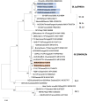
A total of ten 1-2-year-old rabbits died within 2 weeks at a facility in Ehime prefecture in May 2019. Necropsy revealed liver discoloration and fragility, hemorrhage of some organs and blood coagulation failure. On histopathologic examination, necrotizing hepatitis was a common finding, together with fibrin thrombi in the small vessels and hemorrhage in some organs. Rabbit hemorrhagic disease (RHD) virus gene was detected in liver samples, and viral particles of approximately 32 nm in diameter were found in the cytoplasm of degenerated hepatocytes by electron microscopy. Phylogenetic analysis based on the partial VP60 gene sequence classified it as Lagovirus europaeus GI.2/RHDV2. This is the first confirmed outbreak of RHD caused by globally emerging GI.2/RHDV2 in Japan.
Keywords: Japan; Lagovirus europaeus GI.2/RHDV2; electron microscopy; rabbit hemorrhagic disease (RHD).
Conflict of interest statement
The authors have no conflicts of interest to declare.
Figures


Similar articles
-
Rabbit hemorrhagic disease virus 2, 2010-2023: a review of global detections and affected species.J Vet Diagn Invest. 2024 Sep;36(5):617-637. doi: 10.1177/10406387241260281. Epub 2024 Jun 19. J Vet Diagn Invest. 2024. PMID: 39344909 Free PMC article. Review.
-
Overcoming species barriers: an outbreak of Lagovirus europaeus GI.2/RHDV2 in an isolated population of mountain hares (Lepus timidus).BMC Vet Res. 2018 Nov 26;14(1):367. doi: 10.1186/s12917-018-1694-7. BMC Vet Res. 2018. PMID: 30477499 Free PMC article.
-
Large-scale lagovirus disease outbreaks in European brown hares (Lepus europaeus) in France caused by RHDV2 strains spatially shared with rabbits (Oryctolagus cuniculus).Vet Res. 2017 Oct 28;48(1):70. doi: 10.1186/s13567-017-0473-y. Vet Res. 2017. PMID: 29080562 Free PMC article.
-
Occurrence of Lagovirus europaeus (Rabbit Hemorrhagic Disease Virus) in Domestic Rabbits in Southwestern Poland in 2019: Case Report.Microbiol Spectr. 2022 Dec 21;10(6):e0229822. doi: 10.1128/spectrum.02298-22. Epub 2022 Nov 29. Microbiol Spectr. 2022. PMID: 36445093 Free PMC article.
-
Characterisation of Lagovirus europaeus GI-RHDVs (Rabbit Haemorrhagic Disease Viruses) in Terms of Their Pathogenicity and Immunogenicity.Int J Mol Sci. 2024 May 14;25(10):5342. doi: 10.3390/ijms25105342. Int J Mol Sci. 2024. PMID: 38791380 Free PMC article. Review.
Cited by
-
First detection and molecular characterization of rabbit hemorrhagic disease virus (RHDV) in Algeria.Front Vet Sci. 2023 Aug 31;10:1235123. doi: 10.3389/fvets.2023.1235123. eCollection 2023. Front Vet Sci. 2023. PMID: 37745217 Free PMC article.
-
Rabbit hemorrhagic disease virus 2, 2010-2023: a review of global detections and affected species.J Vet Diagn Invest. 2024 Sep;36(5):617-637. doi: 10.1177/10406387241260281. Epub 2024 Jun 19. J Vet Diagn Invest. 2024. PMID: 39344909 Free PMC article. Review.
References
MeSH terms
LinkOut - more resources
Full Text Sources
Other Literature Sources

