

AN ASSOCIATION BETWEEN STELLATE NONHEREDITARY IDIOPATHIC FOVEOMACULAR RETINOSCHISIS, PERIPHERAL RETINOSCHISIS, AND POSTERIOR HYALOID ATTACHMENT
- PMID: 33840784
- PMCID: PMC7611880
- DOI: 10.1097/IAE.0000000000003191
AN ASSOCIATION BETWEEN STELLATE NONHEREDITARY IDIOPATHIC FOVEOMACULAR RETINOSCHISIS, PERIPHERAL RETINOSCHISIS, AND POSTERIOR HYALOID ATTACHMENT
Abstract
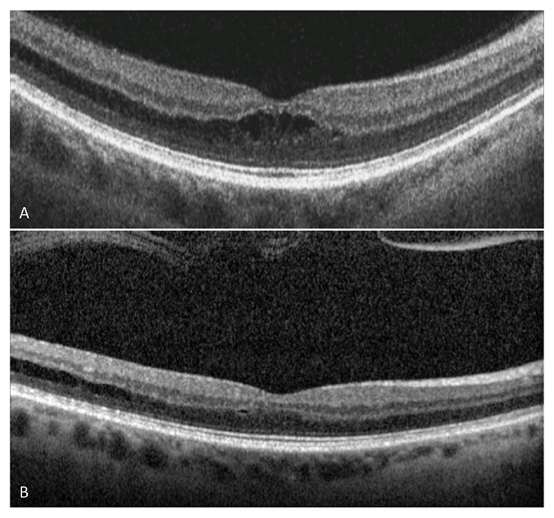
Purpose: Stellate nonhereditary idiopathic foveomacular retinoschisis is a disorder characterized by splitting of the retina at the macula, without a known underlying mechanical or inherited cause. This study investigates demographic, anatomical, and functional characteristics of subjects with stellate nonhereditary idiopathic foveomacular retinoschisis, to explore potential underlying mechanisms.
Methods: In this single-site, retrospective, and cross-sectional, observational study, data were collected from 28 eyes from 24 subjects with stellate nonhereditary idiopathic foveomacular retinoschisis. Descriptive statistics were reported, based on the observed anatomico-functional features.
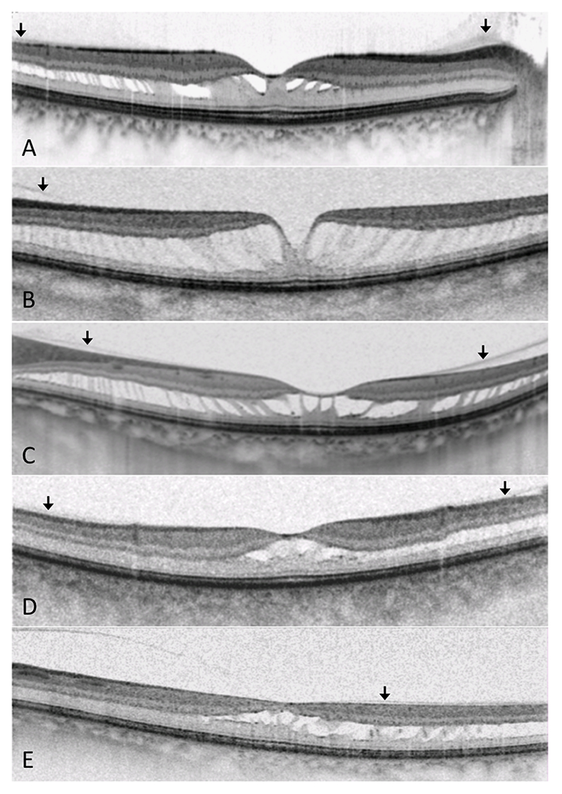
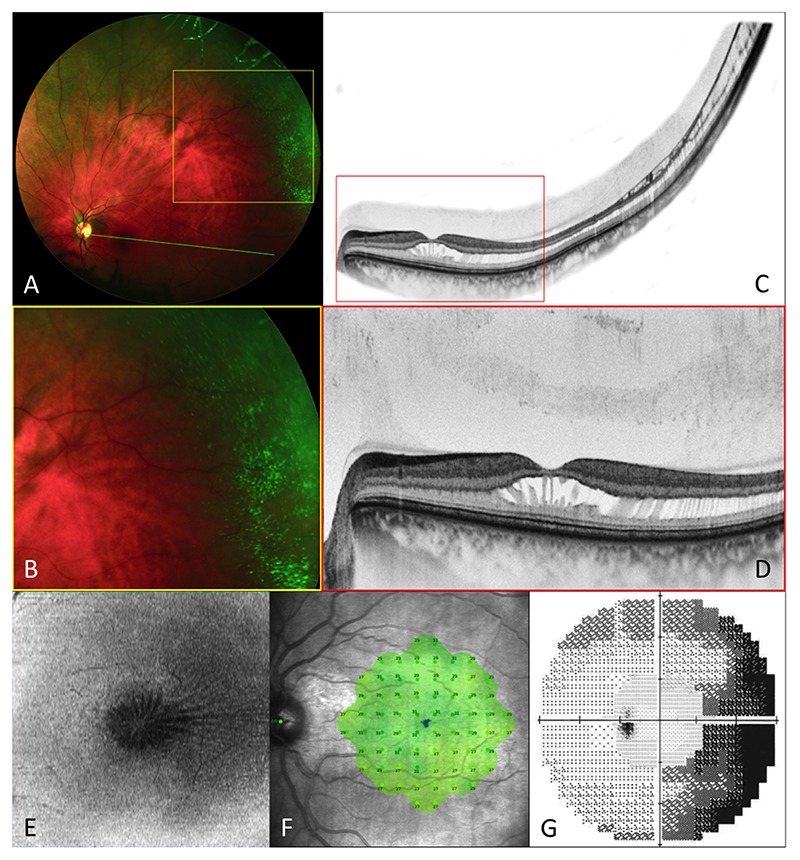
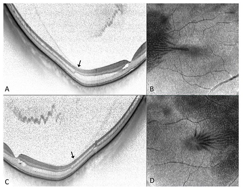
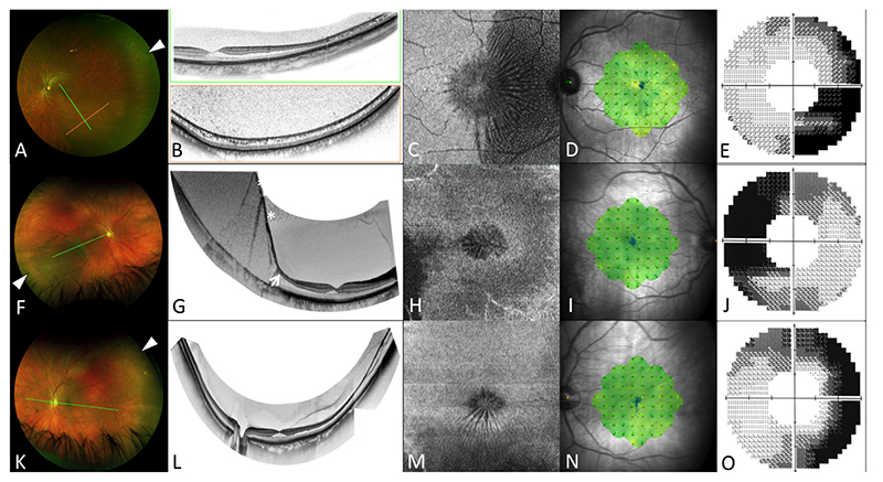
Results: The visual acuity remained stable (median 20/20) in all subjects over a median follow-up of 17 months. All cases demonstrated foveomacular retinoschisis within Henle's fiber layer, at the junction of the outer plexiform and outer nuclear layers. This schisis cavity extended beyond the limits of the macular OCT temporally in all eyes. In most affected eyes, there were documented features of peripheral retinoschisis and broad attachment of the posterior hyaloid at the macula. Functional testing in a cross-sectional subset demonstrated normal retinal sensitivity centrally but an absolute scotoma peripherally.
Conclusion: Stellate nonhereditary idiopathic foveomacular retinoschisis seems to be associated with peripheral retinoschisis and anomalous or incomplete posterior hyaloid detachment. Despite chronic manifestation, this does not significantly affect central visual function but can manifest with profound loss of peripheral visual function.
Copyright © 2021 The Author(s). Published by Wolters Kluwer Health, Inc. on behalf of the Opthalmic Communications Society, Inc.
Figures
References
-
- Yoshida-Uemura T, Katagiri S, Yokoi T, et al. Different foveal schisis patterns in each retinal layer in eyes with hereditary juvenile retinoschisis evaluated by en-face optical coherence tomography. Graefes Arch Clin Exp Ophthalmol. 2017;255(4):719–23. - PubMed
-
- Audo I, Michaelides M, Robson AG, et al. Phenotypic variation in enhanced S-cone syndrome. Invest Ophthalmol Vis Sci. 2008;49(5):2082–93. - PubMed
-
- Bloch E, Georgiadis O, Lukic M, da Cruz L. Optic Disc Pit Maculopathy: New Perspectives on the Natural History. Am J Ophthalmol. 2019;207:159–69. - PubMed
-
- Steel DHW, Suleman J, Murphy DC, et al. Optic disc pit maculopathy: A two-year nationwide prospective population-based study. Ophthalmology. 2018;125(11):1757–64. - PubMed
Publication types
MeSH terms
Grants and funding
LinkOut - more resources
Full Text Sources
Other Literature Sources
Medical
