

Intrinsically Photosensitive Retinal Ganglion Cells of the Human Retina
- PMID: 33841306
- PMCID: PMC8027232
- DOI: 10.3389/fneur.2021.636330
Intrinsically Photosensitive Retinal Ganglion Cells of the Human Retina
Abstract
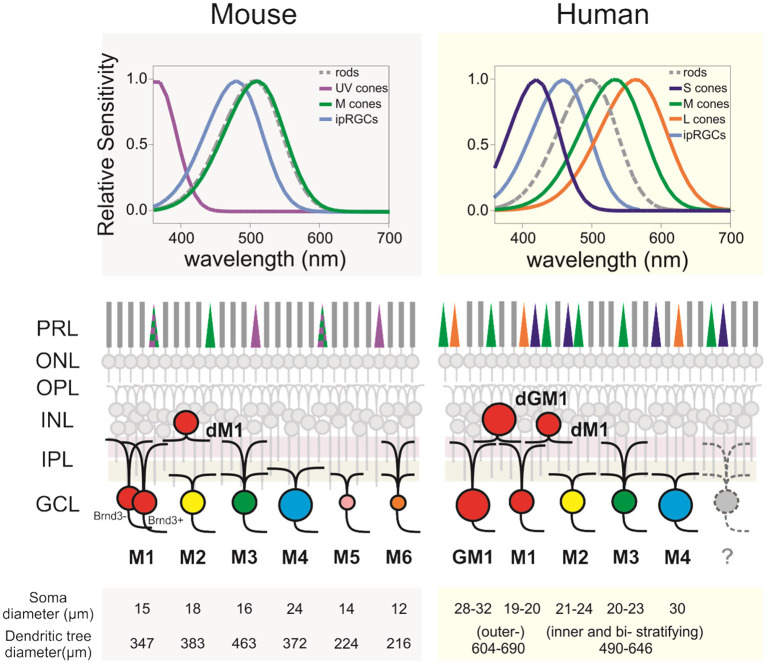
Light profoundly affects our mental and physical health. In particular, light, when not delivered at the appropriate time, may have detrimental effects. In mammals, light is perceived not only by rods and cones but also by a subset of retinal ganglion cells that express the photopigment melanopsin that renders them intrinsically photosensitive (ipRGCs). ipRGCs participate in contrast detection and play critical roles in non-image-forming vision, a set of light responses that include circadian entrainment, pupillary light reflex (PLR), and the modulation of sleep/alertness, and mood. ipRGCs are also found in the human retina, and their response to light has been characterized indirectly through the suppression of nocturnal melatonin and PLR. However, until recently, human ipRGCs had rarely been investigated directly. This gap is progressively being filled as, over the last years, an increasing number of studies provided descriptions of their morphology, responses to light, and gene expression. Here, I review the progress in our knowledge of human ipRGCs, in particular, the different morphological and functional subtypes described so far and how they match the murine subtypes. I also highlight questions that remain to be addressed. Investigating ipRGCs is critical as these few cells play a major role in our well-being. Additionally, as ipRGCs display increased vulnerability or resilience to certain disorders compared to conventional RGCs, a deeper knowledge of their function could help identify therapeutic approaches or develop diagnostic tools. Overall, a better understanding of how light is perceived by the human eye will help deliver precise light usage recommendations and implement light-based therapeutic interventions to improve cognitive performance, mood, and life quality.
Keywords: intrinsically photosensitive ganglion cell; melanopsin (OPN4); non-visual responses to light; retina; retinal ganglion cell.
Copyright © 2021 Mure.
Conflict of interest statement
The author declares that the research was conducted in the absence of any commercial or financial relationships that could be construed as a potential conflict of interest.
Figures
Similar articles
-
The Roles of Rods, Cones, and Melanopsin in Photoresponses of M4 Intrinsically Photosensitive Retinal Ganglion Cells (ipRGCs) and Optokinetic Visual Behavior.Front Cell Neurosci. 2018 Jul 12;12:203. doi: 10.3389/fncel.2018.00203. eCollection 2018. Front Cell Neurosci. 2018. PMID: 30050414 Free PMC article.
-
M1 ipRGCs Influence Visual Function through Retrograde Signaling in the Retina.J Neurosci. 2016 Jul 6;36(27):7184-97. doi: 10.1523/JNEUROSCI.3500-15.2016. J Neurosci. 2016. PMID: 27383593 Free PMC article.
-
[Intrinsically photosensitive retinal ganglion cells].Ophthalmologe. 2022 Apr;119(4):358-366. doi: 10.1007/s00347-021-01476-4. Epub 2021 Aug 4. Ophthalmologe. 2022. PMID: 34350494 Free PMC article. Review. German.
-
Adeno-associated virus (AAV)-mediated Cre recombinase expression in melanopsin ganglion cells without leaky expression in rod/cone photoreceptors.J Neurosci Methods. 2023 Jan 15;384:109762. doi: 10.1016/j.jneumeth.2022.109762. Epub 2022 Dec 5. J Neurosci Methods. 2023. PMID: 36470470 Free PMC article.
-
Burning the candle at both ends: Intraretinal signaling of intrinsically photosensitive retinal ganglion cells.Front Cell Neurosci. 2023 Jan 6;16:1095787. doi: 10.3389/fncel.2022.1095787. eCollection 2022. Front Cell Neurosci. 2023. PMID: 36687522 Free PMC article. Review.
Cited by
-
Circadian regulation in aging: Implications for spaceflight and life on earth.Aging Cell. 2023 Sep;22(9):e13935. doi: 10.1111/acel.13935. Epub 2023 Jul 26. Aging Cell. 2023. PMID: 37493006 Free PMC article. Review.
-
Effects of light therapy on sleep/wakefulness, daily rhythms, and the central orexin system in a diurnal rodent model of seasonal affective disorder.J Affect Disord. 2023 Jul 1;332:299-308. doi: 10.1016/j.jad.2023.04.012. Epub 2023 Apr 13. J Affect Disord. 2023. PMID: 37060954 Free PMC article.
-
Hypersensitivity of Intrinsically Photosensitive Retinal Ganglion Cells in Migraine Induces Cortical Spreading Depression.Int J Mol Sci. 2024 Jul 22;25(14):7980. doi: 10.3390/ijms25147980. Int J Mol Sci. 2024. PMID: 39063222 Free PMC article.
-
Brain stimulation with 40 Hz heterochromatic flicker extended beyond red, green, and blue.Sci Rep. 2024 Jan 25;14(1):2147. doi: 10.1038/s41598-024-52679-z. Sci Rep. 2024. PMID: 38273009 Free PMC article.
-
Ecological factors are likely drivers of eye shape and colour pattern variations across anthropoid primates.Sci Rep. 2022 Oct 15;12(1):17240. doi: 10.1038/s41598-022-20900-6. Sci Rep. 2022. PMID: 36243745 Free PMC article.
References
Publication types
LinkOut - more resources
Full Text Sources
Other Literature Sources
Miscellaneous