. 2021 Mar 15;13(3):988-1005.
eCollection 2021.

Synergistic anti-tumor effect of anti-PD-L1 antibody cationic microbubbles for delivery of the miR-34a gene combined with ultrasound on cervical carcinoma

Affiliations

Synergistic anti-tumor effect of anti-PD-L1 antibody cationic microbubbles for delivery of the miR-34a gene combined with ultrasound on cervical carcinoma

Yun Liu et al. Am J Transl Res. .

Abstract

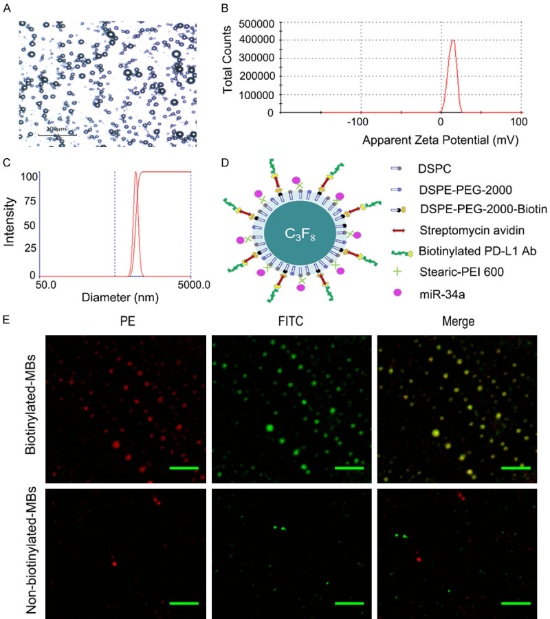
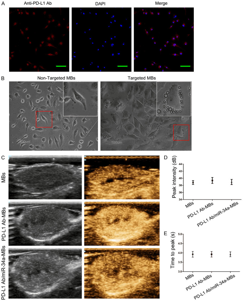
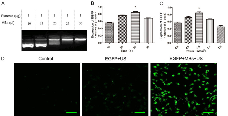
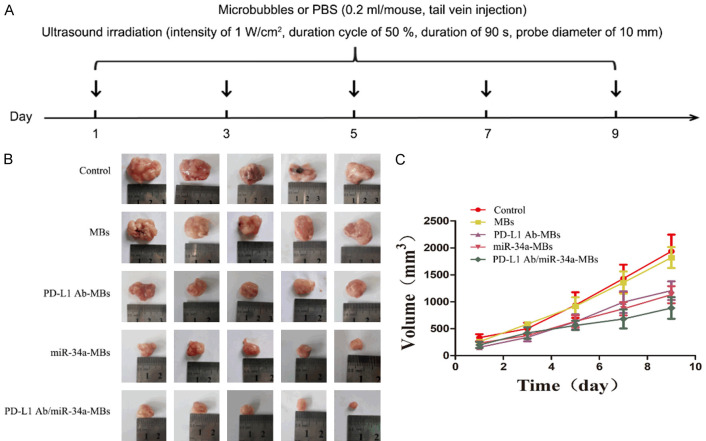
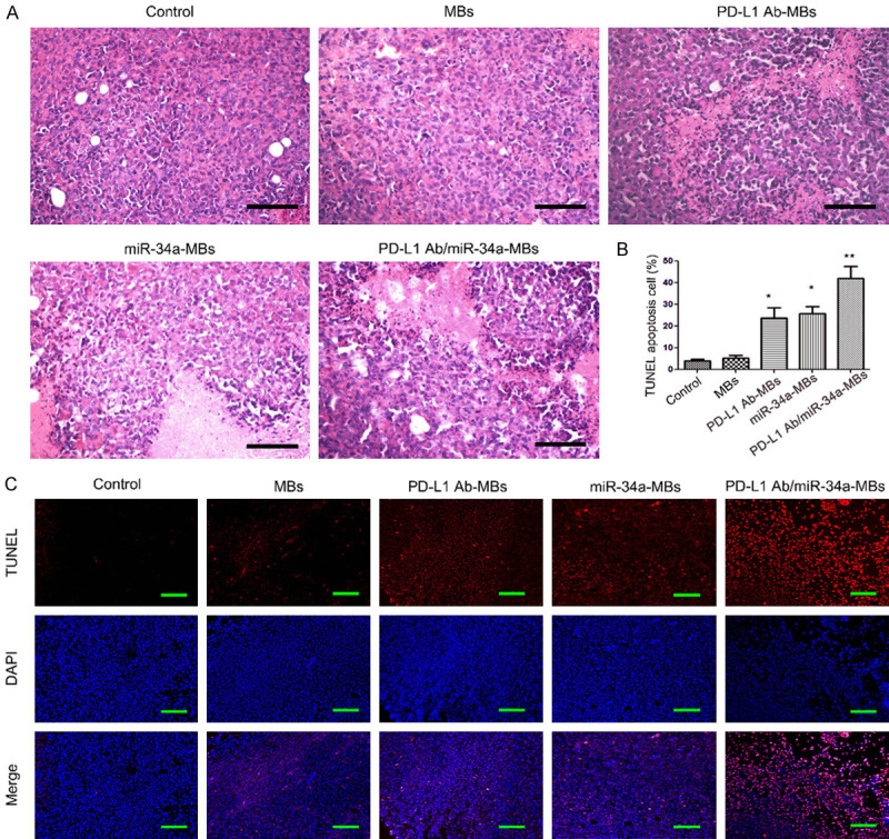
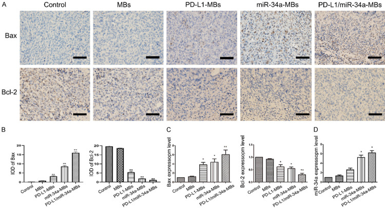
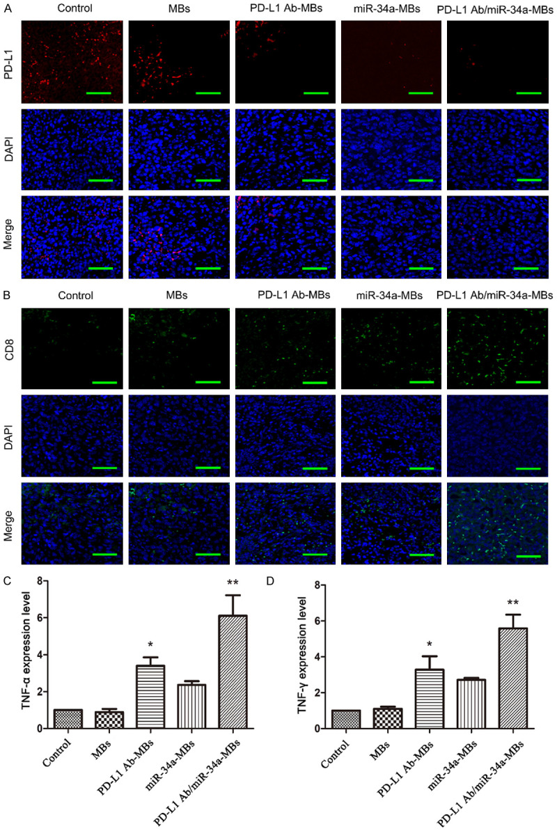
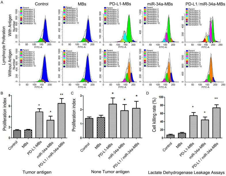
This study explored the synergistic effect of anti-PD-L1 antibody cationic microbubbles (MBs) for delivery of the miR-34a gene combined with ultrasound in inhibiting the cervical cancer. H&E stain, TUNEL, immunohistochemistry and RT-PCR were used to detect the change of apoptosis regulatory factors, and immunofluorescence, Flow cytometry and LDH assays were applied to evaluate the changing of immunomodulatory. In this experiment the PD-L1 Ab/miR-34a-MBs were prepared successfully. The cell targeting assay showed that U14 cells were surrounded by the PD-L1 Ab/miR-34a-MBs and microbubbles had well contrast imaging capability in vivo. With the irradiation power was 1 W/cm2 and the irradiation time was 25 s, the gene transfection efficiency was the highest using EGFP plasmid lorded microbubbles. In vivo anti-tumor assays, the PD-L1 Ab/miR-34a-MBs showed a great potential in inhibiting tumor growth with a TGI of >50%. PD-L1 Ab/miR-34a-MBs treatment enhanced the anti-tumor effect compared with that induced by PD-L1 Ab or miR-34a alone. Firstly, PD-L1 Ab/miR-34a-MBs could gather miR-34a with high-concentration aggregation and releasing around the cervical cancer, which takes a significant role in promoting apoptosis by downregulated Bcl-2 and upregulated Bax. Furthermore, combination therapy was found to augment the activation of T lymphocytes proliferation and increase CD8+ T cells infiltration, to enhance antitumor immune killing effect. The anti-PD-L1 antibody microbubbles for delivery miR-34a gene with ultrasound were considered to be a promising combination therapy regimen via initiating apoptotic mechanism of the tumor and anti-tumor immune regulation.

Keywords: Ultrasound; anti-PD-L1 antibody; cervical cancer; miR-34a; microbubbles.


Conflict of interest statement

None.

Figures

Figure 1
Figure 1
Identification of the prepared targeted microbubbles. A. Characterization of cationic microbubbles were observed by optical microscope (×400); B. The apparent zeta potential of cationic microbubbles was 17.7±3.8 mV. C. The cationic microbubbles had a size distribution of 938.9±80 nm; D. PD-L1 Ab/miR-34a MBs structure diagram; E. Observed PD-L1-Ab conjugation to biotinylated microbubbles and non-biotinylated microbubbles by laser confocal microscopy (PE: streptavidin-PE; FITC: rabbit anti-mouse IgG-Fc/FITC) (×400), scale bar = 50 μm. Red fluorescence indicates that streptavidin-PE binds to the biotin of the microbubble, and green fluorescence indicates that the FITC-labeled secondary antibody binds to the mouse anti-PD-L1 antibody and then links with the microbubbles.
Figure 2
Figure 2
Examined the targeting and contrast-enhanced efficiency of targeted-MBs in intro and vivo. (A) PD-L1 expression on U14 membranes was confirmed by laser confocal microscopy. High expression of red fluorescence was observed on the U14 membranes (×200), scale bar = 100 μm; (B) Non-targeted microbubbles (NMBs) and targeted microbubbles (TMBs) were observed bind to U14 cells by light microscope (×400), with NMBs, only a slight non-specificity binding to U14s was observed; comparatively, it can be observed that the U14 cells were surrounded by TMBs; (C) The results of Contrast-enhanced Ultrasound Imaging showed that all of the MBs, PD-L1 Ab-MBs and PD-L1 Ab/miR-34a-MBs had well contrast imaging capabilities in vivo. The peak intensity (D) and the time to peak (E) of Contrast-enhanced Ultrasound Imaging in vivo have no significant difference among the groups (P>0.05). Date in figure D&E are presented as mean ± SD, n = 3.
Figure 3
Figure 3
The UTMD promoted gene transfection into U14 cells through the DNA loaded targeted microbubbles. A. Analysis of DNA loading capacity of the MBs. The result of agarose gel electrophoresis (AGE) showed that the 25 μl MBs has the capacity of loading 1 μg plasmids. B, C. Quantitative assessment of UTMD-mediated gene transfection in U14 cell with different irradiation power and time; D. The fluorescence microscopic images showed that, control group and EGFP pasmid + US group exhibit negligible fluorescence emission, while, EGFP plasmid-MBs + US group exhibited the higher fluorescence emission (×200), scale bar = 100 μm.
Figure 4
Figure 4
The in vivo therapeutic effects of the targeted microbubbles in the U14 cervical carcinoma bearing mice. A. Schematic illustration of the therapeutic formulations. Mice were injected with different types of microbubbles, while in control group were injected with equal dosage of PBS, and each group was irradiated by ultrasound with specific parameters every two days (n = 10). B. Representative tumor tissues excised from the mice at the end of the treatment; C. Tumor volume growth curve. The tumor volume of the control group and MBs + US group showed an exponential growth pattern, and the tumor growth of the other groups was slowed down.
Figure 5
Figure 5
Analysis of the apoptosis of tumor tissues after the treatment. (A) Histopathology of tumor tissues in each group (H&E, ×400), scale bar = 200 μm. The histological sections were prepared from various tumor tissues in the mice. In the control group or MBs + US group the tumor histopathological sections showed that the arrangement of tumor cells was disordered, tumor cells active growth and evident nucleic division, and tumor cells with large nuclear-cytoplasmic ratios and significant profile; While in the targeted MBs the tumor histopathological sections revealed that the tumor cells extensive necrosis, especially in the PD-L1 Ab/miR-34a-MBs + US group; (B) The semi-quantification analysis of apoptotic cells in tumor tissues of each group by the fluorescence intensity showed in (C) (Compared with the Control group, *P<0.05, **P<0.01). (C) TUNEL assay was used to analyse cell apoptosis. Red-stained nuclei indicate cells in the apoptotic state (original magnification, ×200), scale bar = 100 μm.
Figure 6
Figure 6
Expression of Bcl-2 and Bax in the tumor tissues. (A) Detection of Bax and Bcl-2 protein expression in tumor tissues by immunohistochemistry staining (×400), scale bar = 200 μm. (B) Data were processed using ImageJ software. The protein expression of Bax shown in (A) was significantly higher in the PD-L1 Ab-MBs + US group, miR-34a-MBs + US group and PD-L1 Ab/miR-34a-MBs + US group, while the results of Bcl-2 expression were reversed. (C) The mRNA expression of Bax and Bcl-2. The expression of Bax gene in all the treatment groups was higher than that in the control groups, the PD-L1 Ab/miR-34a-MBs + US group was most obvious. While the results of Bcl-2 were reversed. (D) The mRNA expression of miR-34a. The expression of miR-34a gene were up-regulated in the treatment groups, especially in the miR-34a-MBs + US group and PD-L1 Ab/miR-34a-MBs + US group. (Compared with the control group, *P<0.05; **P<0.01, n = 10).
Figure 7
Figure 7
The anti-tumor immunotherapy with the targeted microbubbles. A. Expression of PD-L1 in each group detected by immunofluorescence (original magnification, ×600), scale bar = 50 μm. The expression of PD-L1 was significantly downregulated in the PD-L1 Ab/miR-34a-MBs + US group compared to Control group. B. Expression of CD8-positive T cells in each group detected by immunofluorescence (original magnification, ×600), scale bar = 50 μm. In addition to the Control group, the number of CD8 T cells in each group of tumor tissues increased, and green fluorescence was emitted significant in PD-L1 Ab/miR-34a-MBs + US group under laser confocal microscope. C, D. The mRNA expression of IFN-γ and TNF-α. The expression of IFN-γ and TNF-α gene in all the treatment groups was higher than that in the control groups, the increased in PD-L1 Ab/miR-34a-MBs + US group was most obvious. (Compared with the Control group, *P<0.05; **P<0.01, n = 10).
Figure 8
Figure 8
Analysis of the spleen lymphocyte proliferation after the targeted microbubbles by flow cytometry. A. Splenocytes were labeled with CFSE and cultured for 72 (with or without tumor antigen stimulation). B. Calculated the lymphocyte proliferation index in each group with tumor antigen; C. Calculated the lymphocyte proliferation index in each group without tumor antigen. D. The cytotoxicity of lymphocytes detected by Lactate dehydrogenase leakage assays, the cytotoxic cell killing rate increased significant in PD-L1 Ab-MBs + US group and miR-34a-MBs + US group, and the increasing of cytotoxic cell killing rate was more significant in PD-L1 Ab/miR-34a-MBs + US group. (Compared with the control group, *P<0.05, **P<0.01, n = 5).

Similar articles

Cited by

References

    1. Bray F, Ferlay J, Soerjomataram I, Siegel RL, Torre LA, Jemal A. Global cancer statistics 2018: GLOBOCAN estimates of incidence and mortality worldwide for 36 cancers in 185 countries. CA Cancer J Clin. 2018;68:394–424. - PubMed
    1. Couzin-Frankel J. Breakthrough of the year 2013. Cancer immunotherapy. Science. 2013;342:1432–1433. - PubMed
    1. Ribas A, Wolchok JD. Cancer immunotherapy using checkpoint blockade. Science. 2018;359:1350–1355. - PMC - PubMed
    1. Fesnak AD, June CH, Levine BL. Engineered T cells: the promise and challenges of cancer immunotherapy. Nat Rev Cancer. 2016;16:566–581. - PMC - PubMed
    1. Tipanee J, Chai YC, VandenDriessche T, Chuah MK. Preclinical and clinical advances in transposon-based gene therapy. Biosci Rep. 2017;37:BSR20160614. - PMC - PubMed

LinkOut - more resources