. 2020 Aug 27:29:13-22.
doi: 10.1016/j.jare.2020.08.013. eCollection 2021 Mar.

Inhibition of lung cancer by vitamin D depends on downregulation of histidine-rich calcium-binding protein

Affiliations

Inhibition of lung cancer by vitamin D depends on downregulation of histidine-rich calcium-binding protein

Ning Liu et al. J Adv Res. .

Abstract

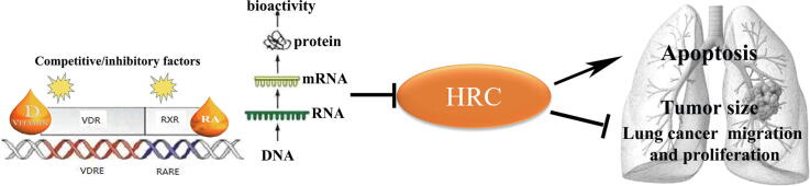
Introduction: Intrinsic vitamin D affects the proliferation, apoptosis, invasion, metastasis, and tumorigenesis of lung cancer by regulating tumor signaling pathways. Histidine-rich calcium-binding protein (HRC) maintains Ca2+ homeostasis, which plays crucial roles in the occurrence and development of cancer.

Objectives: Our study aims to investigate the ability of vitamin D in the regulation of HRC and the role of HRC playing in lung cancer.

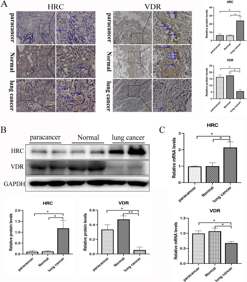
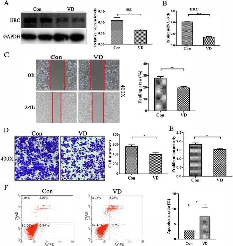
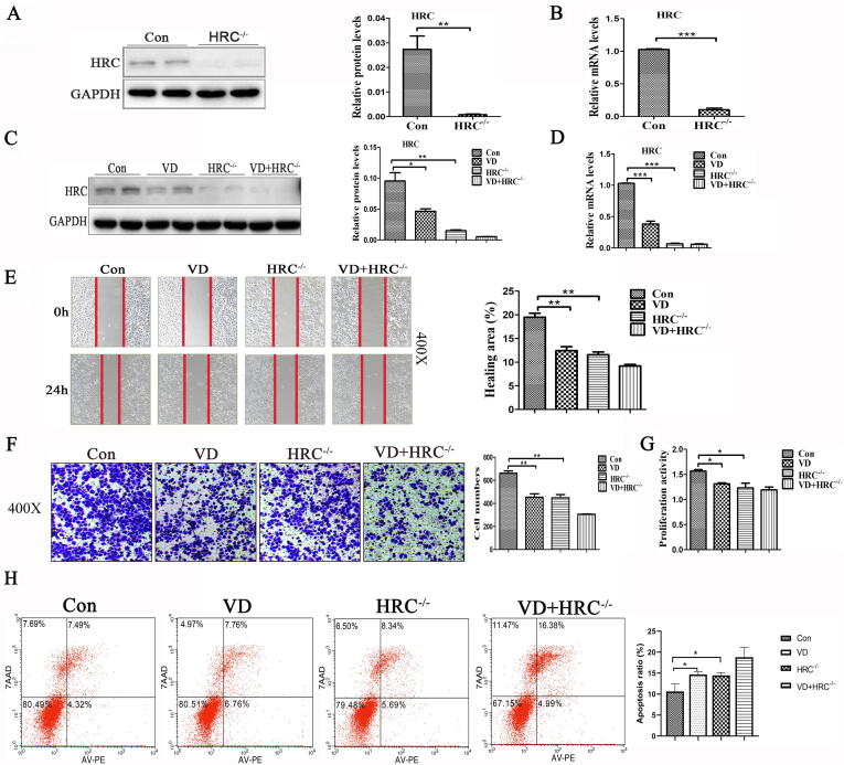
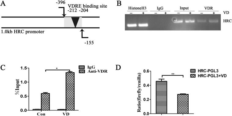
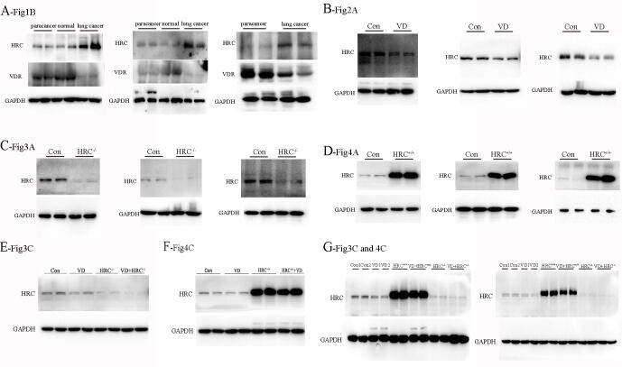
Methods: We investigated the effects of vitamin D on lung cancer and the underlying mechanisms, by measuring HRC and vitamin D receptor (VDR) expression in lung cancer, paracancer, and normal tissues from patients using immunohistochemistry, western blotting, and real time RT-PCR. We transfected H460 lung cancer cells (supplemented or not with vitamin D) with PX458-HRC and pcDNA3.1-HRC plasmids and injected mice with lung cancer cells harboring pcDNA3.1-vector or pcDNA3.1-HRC plasmids.

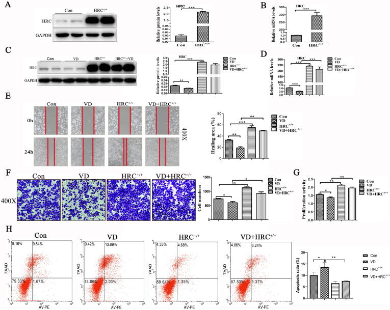
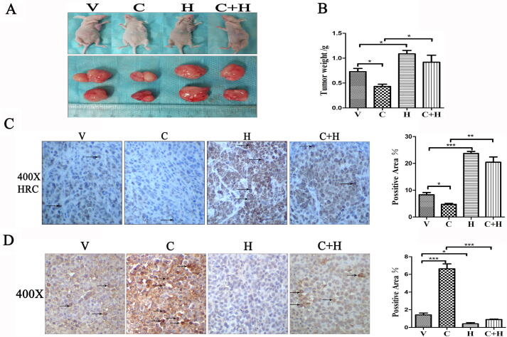
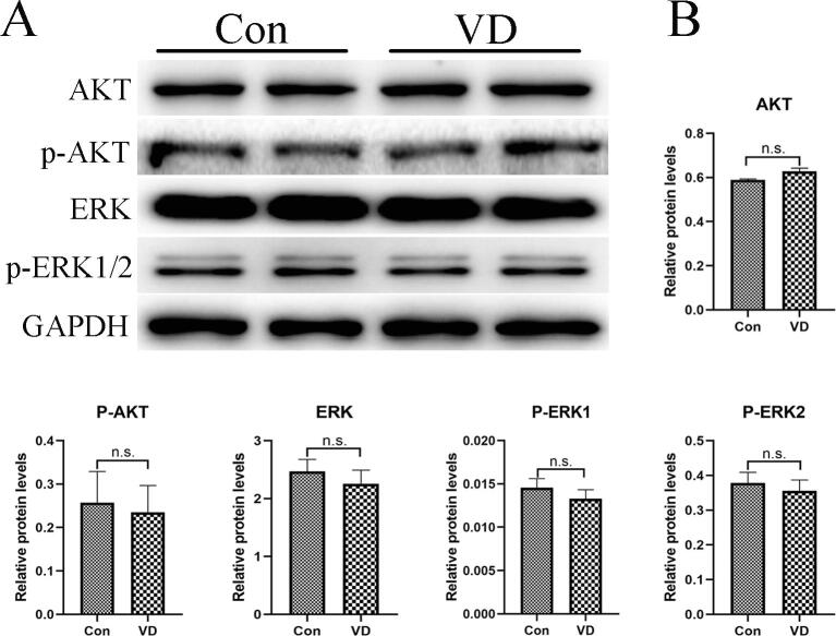
Results: Vitamin D inhibited HRC expression and H460 cell migration and proliferation, and promoted apoptosis compared with controls. The expression of HRC and VDR was significantly upregulated and downregulated, respectively, in lung cancer versus paracancer or normal tissues. Cell proliferation and migration were reduced, apoptotic cells were more and tumors were smaller in mice treated with vitamin D/cholecalciferol cholesterol emulsion (CCE) than in vitamin D/CCE+HRC+/+ mice.

Conclusion: Vitamin D inhibited lung cancer tumor growth, migration, and proliferation by downregulating HRC.

Keywords: Histidine-rich calcium-binding protein; Lung cancer; Vitamin D; Vitamin D receptor.


Conflict of interest statement

The authors declare that they have no known competing financial interests or personal relationships that could have appeared to influence the work reported in this paper.

Figures

None
Graphical abstract
Fig. 1
Fig. 1
Expression of VDR and HRC respectively decreased and increased in lung cancer. (A) Immunohistochemical staining and (B) western blot of HRC and VDR protein expression in lung cancer, paracancer and normal tissues. The expression is shown as relative to GAPDH protein levels determined using Prism 5. Blue arrow, positive area (original magnification, ×200; partial magnification of ×400). (C) Levels of HRC and VDR mRNA expression in lung cancer, paracancer and normal tissues measured using real time TR-PCR. Values are shown as means ± SD. Statistical significance is shown as *P < 0.05, **P < 0.01, ***P < 0.001. HRC, histidine-rich calcium binding protein; VDR, vitamin D receptor. (For interpretation of the references to colour in this figure legend, the reader is referred to the web version of this article.)
Fig. 2
Fig. 2
Vitamin D downregulated HRC expression and inhibited H460 cell growth. (A) Levels of HRC protein expression in H460 cells incubated with or without vitamin D. The histogram shows that vitamin D significantly reduced HRC expression. (B) Real time RT-PCR results show that vitamin D significantly reduced HRC mRNA levels. (C) Wound healing assay. Vitamin D significantly reduced cell mobility, expressed as the ratio (%) of healed areas after 24 h. Red line, edge of cell migration. (D) Transwell migration assays. The histogram shows vitamin D-inhibited cell migration and numbers of migrated cells. (E) Cell proliferation assays. Vitamin D inhibited H460 cell proliferation. (F) Apoptosis induction assessed by flow cytometry after Annexin V-PE/7-AAD staining. Vitamin D promoted apoptosis. Values are expressed as means ± SD. Statistical significance is shown as *P < 0.05, **P < 0.01, and ***P < 0.001. HRC, Histidine-rich calcium binding protein. (For interpretation of the references to colour in this figure legend, the reader is referred to the web version of this article.)
Fig. 3
Fig. 3
Synergistic effect of vitamin D and HRC−/− on cell migration, proliferation and apoptosis. (A) Protein expression of HRC in H460 cell lines with HRC knockdown. Western blots show that HRC has been knocked out. (B) HRC was detected at the level of mRNA by real time RT-PCR. (C) Western blots of HRC expression show that vitamin D and HRC−/− further reduced HRC expression. (D) Levels of HRC mRNA detected by real time RT-PCR. (E) Wound healing assay. Vitamin D and HRC knockdown both reduced cell mobility, but together, the reduction in cell migration was further reduced. (F) Transwell migration assays revealed that vitamin D and HRC−/− synergistically inhibited H460 cell migration. (G) Proliferation of H460 cells. Results of MTS assays showed good synergistic effects. (H) Apoptosis induction assessed by flow cytometry after annexin V-PE/7-AAD staining. Vitamin D and HRC−/− synergistically promoted apoptosis. Values are expressed as means ± SD. Statistical significance is shown as *P < 0.05, **P < 0.01, and ***P < 0.001. HRC, Histidine-rich calcium binding protein.
Fig. 4
Fig. 4
HRC+/+ attenuated the effects of vitamin D on cell migration, proliferation and apoptosis. (A) Western blots show HRC overexpression in H460 cells; a 200-fold increase in HRC expression was noted. (B) Overexpressed HRC detected by real time RT-PCR. (C) The histogram shows HRC protein expression after 24 h incubation with vitamin D. (D) Effects of vitamin D on HRC mRNA expression determined by real time RT-PCR. (E) Wound healing assay. Vitamin D with HRC+/+ partially reduced cell mobility compared with that with HRC+/+, but the effect was weaker than that in the VD group. (F) Transwell migration assays. The histogram shows that vitamin D with HRC+/+ partially inhibited H460 cell migration. (G) Assays of H460 cell proliferation showed that HRC+/+ attenuates the effects of vitamin D on cell migration and proliferation. (H) Apoptosis induction assessed by flow cytometry after staining with annexin V-PE/7-AAD. Vitamin D-induced H460 cell apoptosis was significantly reduced by HRC+/+. Values are expressed as means ± SD. Statistical significance is shown as *P < 0.05, **P < 0.01, and ***P < 0.001. HRC, Histidine-rich calcium binding protein.
Fig. 5
Fig. 5
Tumor growth was partially inhibited in the CCE+HRC group compared with the HRC group in vivo. (A) Images of tumors in the xenograft nude mice. Tumors were the smallest in the CCE group, and tumor reduction by CCE was weakened by HRC. (B) Mean tumor weight of nude mice. Tumor growth was partially inhibited by CCE under HRC overexpression, but the effect was weaker than that under CCE treatment alone. (C) Immunohistochemical staining shows HRC expression among groups. (D) TUNEL staining was more intense in the CCE group than in the CCE+HRC group. Arrow, site of apoptosis. Values are expressed as means ± SD. Statistical significance is shown as *P < 0.05, **P < 0.01, and ***P < 0.001. CCE, cholecalciferol cholesterol emulsion; HRC, histidine-rich calcium binding protein; TUNEL, terminal deoxynucleotidyl transferase-mediated dUTP nick-end labeling.
Fig. 6
Fig. 6
HRC is the direct target of vitamin D. (A) Schema of the HRC promoter region. (B) ChIP assays of H460 cells using anti-VDR antibody. Vitamin D response elements in the HRC promoter amplified by PCR. (C) Immunoprecipitated DNA with protein detected by ChIP-qPCR. The bar graph shows the ratio (%) of relative fold enrichment of VDR across the HRC promoter region. (D) Cos-7 cells transfected with HRC-PGL3 reporter plasmid. Vitamin D reduced the transcriptional activity of HRC. Values are expressed as means ± SD. Statistical significance is shown as *P < 0.05 and **P < 0.01. HRC, histidine-rich calcium binding protein; VDR, vitamin D receptor.
Supplementary fig. 1
Replicates of all western blots have been shown in this figure. Replicates of (A) fig.1B, (B) fig.2A, (C) fig.3A, (D) fig.4A, (E) fig.3C, (F) fig.4C, and (G) fig.3C and 4C.
Supplementary fig. 2
Vitamin D does not regulate the protein expression of AKT, p-AKT, ERK and p-ERK1/2 in H460 cells. (A) Western blots show that levels of AKT, P-AKT, ERK and P-ERK protein expression in H460 cells incubated with or without vitamin D. (B) The histograms show that the differences between the Con and VD groups are not statistically significant. Values are expressed as means ± SD. Statistical significance is shown as n.s. (P >0.05, not significant). AKT, protein kinase B; p-AKT, phospho-protein kinase B; ERK; extracellular regulated protein kinases; p-ERK, phospho-extracellular regulated protein kinases.

Similar articles

Cited by

References

    1. Nasim F., Sabath B.F., Eapen G.A. Lung cancer. Med Clin North Am. 2019;103:463–473. doi: 10.1016/j.mcna.2018.12.006. - DOI - PubMed
    1. Maj E., Filip-Psurska B., Milczarek M., Psurski M., Kutner A., Wietrzyk J. Vitamin D derivatives potentiate the anticancer and anti-angiogenic activity of tyrosine kinase inhibitors in combination with cytostatic drugs in an A549 non-small cell lung cancer model. Int J Oncol. 2018;52:337–366. doi: 10.3892/ijo.2017.4228. - DOI - PMC - PubMed
    1. Picello E., Damiani E., Margreth A. Low-affinity Ca(2+)-binding sites versus Zn(2+)-binding sites in histidine-rich Ca(2+)-binding protein of skeletal muscle sarcoplasmic reticulum. Biochem Biophys Res Commun. 1992;186:659–667. - PubMed
    1. Arvanitis D.A., Vafiadaki E., Johnson D.M., Kranias E.G., Sanoudou D. The histidine-rich calcium binding protein in regulation of cardiac rhythmicity. Front Physiol. 2018;9:1379. doi: 10.3389/fphys.2018.01379. - DOI - PMC - PubMed
    1. Arvanitis D.A., Vafiadaki E., Fan G.C., Mitton B.A., Gregory K.N., Del Monte F. Histidine-rich Ca-binding protein interacts with sarcoplasmic reticulum Ca-ATPase. Am J Physiol Heart Circulatory Physiol. 2007;293:H1581–H1589. doi: 10.1152/ajpheart.00278.2007. - DOI - PubMed

Publication types

MeSH terms