

Comparison of tear proteomic and neuromediator profiles changes between small incision lenticule extraction (SMILE) and femtosecond laser-assisted in-situ keratomileusis (LASIK)
- PMID: 33842006
- PMCID: PMC8020296
- DOI: 10.1016/j.jare.2020.11.001
Comparison of tear proteomic and neuromediator profiles changes between small incision lenticule extraction (SMILE) and femtosecond laser-assisted in-situ keratomileusis (LASIK)
Abstract
Introduction: The tear proteomics and neuromediators are associated with clinical dry eye parameters following refractive surgery.
Purpose: To investigate and compare the tear proteomic and neuromediator profiles following small incision lenticule extraction (SMILE) versus laser-assisted in-situ keratomileusis (LASIK).
Methods: In this randomized controlled trial with paired-eye design, 70 patients were randomized to receive SMILE in one eye and LASIK in the other eye. Tear samples were collected preoperatively, and 1 week, 1, 3, 6 and 12 months postoperatively, and were examined for protein concentration changes using sequential window acquisition of all theoretical fragment ion mass spectrometry (SWATH-MS). The data were analyzed with DAVID Bioinformatics Resources for enriched gene ontology terms and over-represented pathways. Tear neuromediators levels were correlated with clinical parameters.
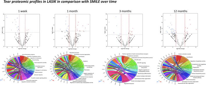
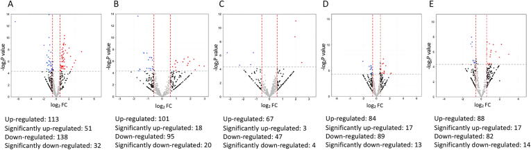
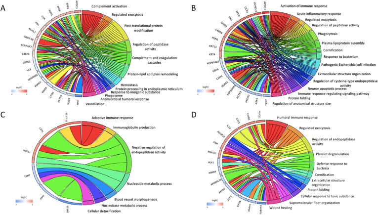
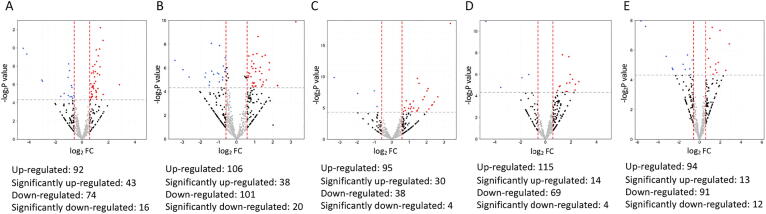
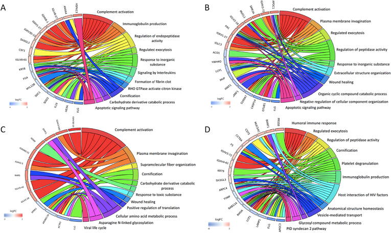
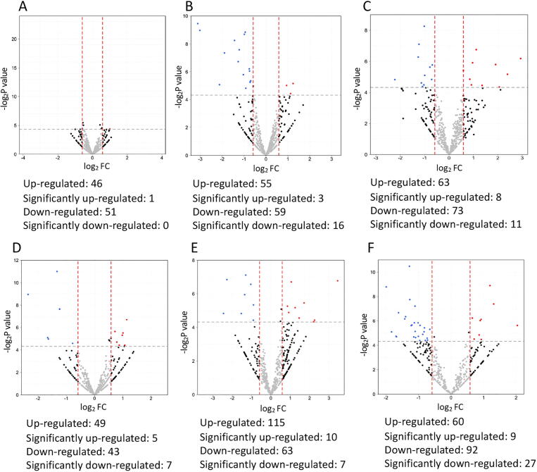
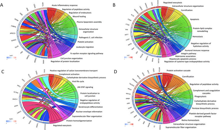
Results: Post-SMILE eyes had significantly better Oxford staining scores and tear break-up time (TBUT) than post-LASIK eyes at 1 and 3 months, respectively. Tear substance P and nerve growth factor levels were significantly higher in the LASIK group for 3 months and 1 year, respectively. SMILE and LASIK shared some similar biological responses postoperatively, but there was significant up-regulation in leukocyte migration and wound healing at 1 week, humoral immune response and apoptosis at 1 month, negative regulation of endopeptidase activity at 3 to 6 months, and extracellular structure organization at 1 year in the post-LASIK eyes. Tear mucin-like protein 1 and substance P levels were significantly correlated with TBUT (r = -0.47, r = -0.49, respectively).
Conclusion: Significant differences in the tear neuromediators and proteomics were observed between SMILE and LASIK, even though clinical dry eye signs have subsided and became comparable between 2 procedures.
Keywords: Dry eye; Laser-assisted in-situ keratomileusis; Neuromediators; Proteomics; Small incision lenticule extraction; Tear.
© 2021 The Authors. Published by Elsevier B.V. on behalf of Cairo University.
Conflict of interest statement
The authors declare that they have no known competing financial interests or personal relationships that could have appeared to influence the work reported in this paper.
Figures
Similar articles
-
Dry Eye after Small Incision Lenticule Extraction (SMILE) versus Femtosecond Laser-Assisted in Situ Keratomileusis (FS-LASIK) for Myopia: A Meta-Analysis.PLoS One. 2016 Dec 16;11(12):e0168081. doi: 10.1371/journal.pone.0168081. eCollection 2016. PLoS One. 2016. PMID: 27992482 Free PMC article. Review.
-
Dry eye disease after refractive surgery: comparative outcomes of small incision lenticule extraction versus LASIK.Ophthalmology. 2015 Apr;122(4):669-76. doi: 10.1016/j.ophtha.2014.10.004. Epub 2014 Nov 22. Ophthalmology. 2015. PMID: 25458707
-
Comparison of Corneal Biological Healing After Femtosecond LASIK and Small Incision Lenticule Extraction Procedure.Curr Eye Res. 2016 Sep;41(9):1202-8. doi: 10.3109/02713683.2015.1107590. Epub 2016 Feb 1. Curr Eye Res. 2016. PMID: 26833247 Clinical Trial.
-
Longitudinal analysis of wound healing response post SMILE and LASIK surgery using proteomic profiling of tears.Exp Eye Res. 2024 Sep;246:109987. doi: 10.1016/j.exer.2024.109987. Epub 2024 Jul 2. Exp Eye Res. 2024. PMID: 38964497
-
Dry Eye After Small Incision Lenticule Extraction and Femtosecond Laser-Assisted LASIK: Meta-Analysis.Cornea. 2017 Jan;36(1):85-91. doi: 10.1097/ICO.0000000000000999. Cornea. 2017. PMID: 27560032 Review.
Cited by
-
Postoperative efficacy, safety, predictability, and visual quality of implantable collamer lens implantation versus small incision lenticule extraction in myopic eyes: a Meta-analysis.Int J Ophthalmol. 2023 Mar 18;16(3):442-452. doi: 10.18240/ijo.2023.03.16. eCollection 2023. Int J Ophthalmol. 2023. PMID: 36935780 Free PMC article.
-
Impact of Dry Eye Disease on the Uncorrected Distance Visual Acuity after Small Incision Lenticule Extraction.J Clin Med. 2023 Sep 25;12(19):6179. doi: 10.3390/jcm12196179. J Clin Med. 2023. PMID: 37834823 Free PMC article.
-
Interconnections between diabetic corneal neuropathy and diabetic retinopathy: diagnostic and therapeutic implications.Neural Regen Res. 2025 Aug 1;20(8):2169-2180. doi: 10.4103/NRR.NRR-D-24-00509. Epub 2024 Sep 20. Neural Regen Res. 2025. PMID: 39359077 Free PMC article.
-
In Vivo Confocal Microscopy Evaluation in Patients with Keratoconus.J Clin Med. 2022 Jan 13;11(2):393. doi: 10.3390/jcm11020393. J Clin Med. 2022. PMID: 35054085 Free PMC article. Review.
-
Chronic Kidney Disease Has No Impact on Tear Film Substance P Concentration in Type 2 Diabetes.Biomedicines. 2023 Aug 24;11(9):2368. doi: 10.3390/biomedicines11092368. Biomedicines. 2023. PMID: 37760810 Free PMC article.
References
-
- Liu Y.C., Rosman M., Mehta J.S. Enhancement after small-incision lenticule extraction: Incidence, risk factors, and outcomes. Ophthalmology. 2017;124(6):813–821. - PubMed
-
- Kobashi H., Kamiya K., Shimizu K. Dry eye after small incision lenticule extraction and femtosecond laser-assisted LASIK: Meta-analysis. Cornea. 2017;36(1):85–91. - PubMed
-
- Denoyer A., Landman E., Trinh L., Faure J.F., Auclin F., Baudouin C. Dry eye disease after refractive surgery: comparative outcomes of small incision lenticule extraction versus LASIK. Ophthalmology. 2015;122(4):669–676. - PubMed
-
- Fuest M., Mehta J.S. Iatrogenic dry eye following cataract and refractive surgical procedures. In: He M.G., editor. Ocular surface diseases, disorders, & dysfunctions. Thorofare; NJ, Healio: 2016. pp. 3–6.
-
- Liu Y.C., Tan D.T.H., Mehta J.S. Wound healing after ReLEx surgery. In: Sekundo W., editor. small incision lenticule extraction: Principles, techniques, complication management and future concepts. NY, Springer; New York: 2015. pp. 13–26.
Publication types
MeSH terms
Substances
LinkOut - more resources
Full Text Sources
Other Literature Sources
Medical
Research Materials
