. 2020 Oct 8:29:95-106.
doi: 10.1016/j.jare.2020.09.009. eCollection 2021 Mar.

A proline-rich motif in the large intracellular loop of the glycine receptor α1 subunit interacts with the Pleckstrin homology domain of collybistin

Affiliations

A proline-rich motif in the large intracellular loop of the glycine receptor α1 subunit interacts with the Pleckstrin homology domain of collybistin

Ulrike Breitinger et al. J Adv Res. .

Abstract

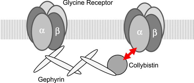
Introduction: The inhibitory glycine receptor (GlyR), a mediator of fast synaptic inhibition, is located and held at neuronal synapses through the anchoring proteins gephyrin and collybistin. Stable localization of neurotransmitter receptors is essential for synaptic function. In case of GlyRs, only beta subunits were known until now to mediate synaptic anchoring.

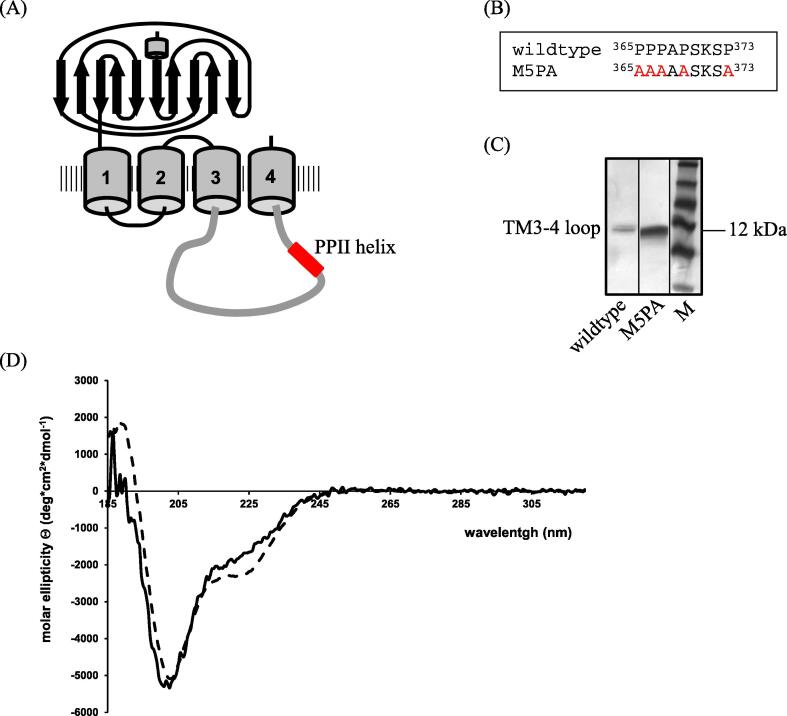
Objectives: We identified a poly-proline II helix (PPII) in position 365-373 of the intra-cellular TM3-4 loop of the human GlyRα1 subunit as a novel potential synaptic anchoring site. The potential role of the PPII helix as synaptic anchoring site was tested.

Methods: Glycine receptors and collybistin variants were generated and recombinantly expressed in HEK293 cells and cultured neurons. Receptor function was assessed using patch-clamp electrophysiology, protein-protein interaction was studied using co-immuno-precipitation and pulldown experiments.

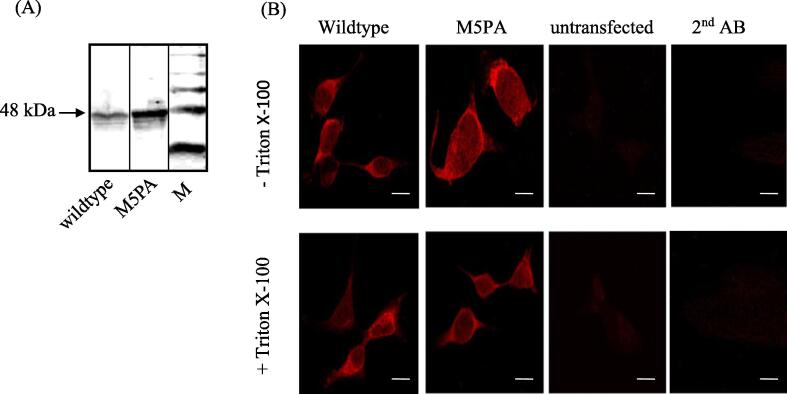
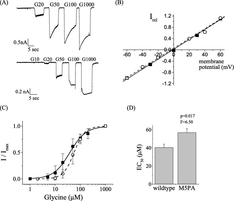
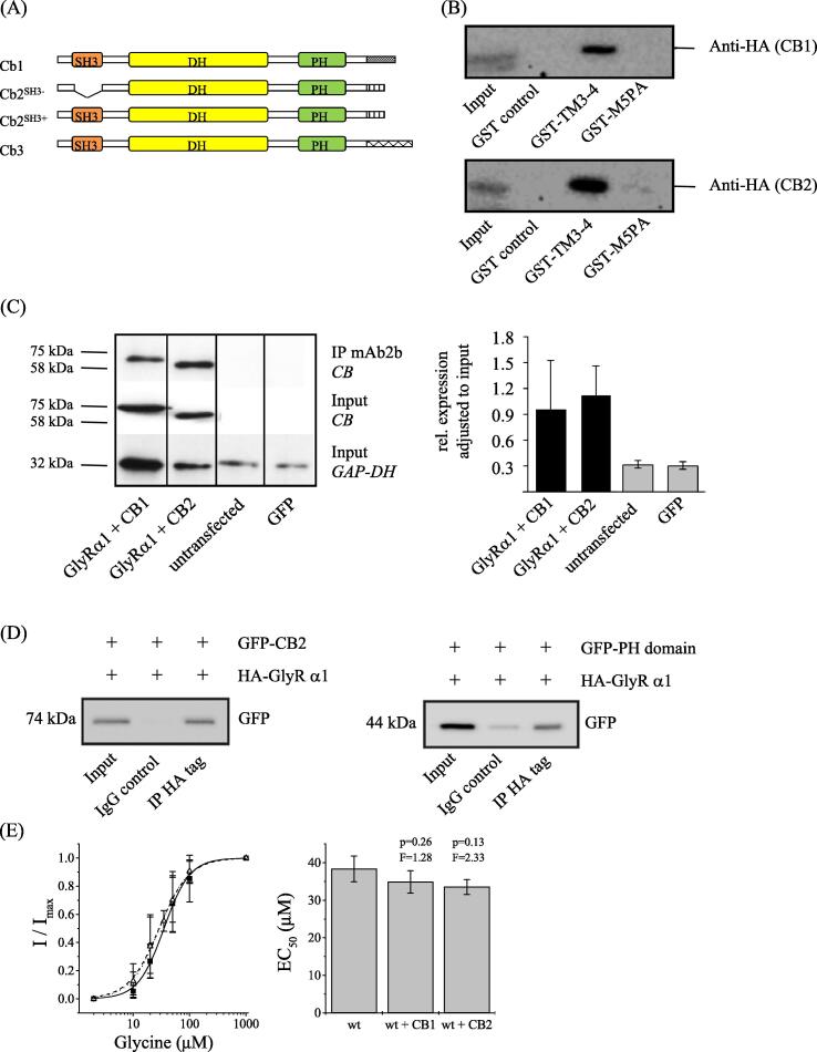
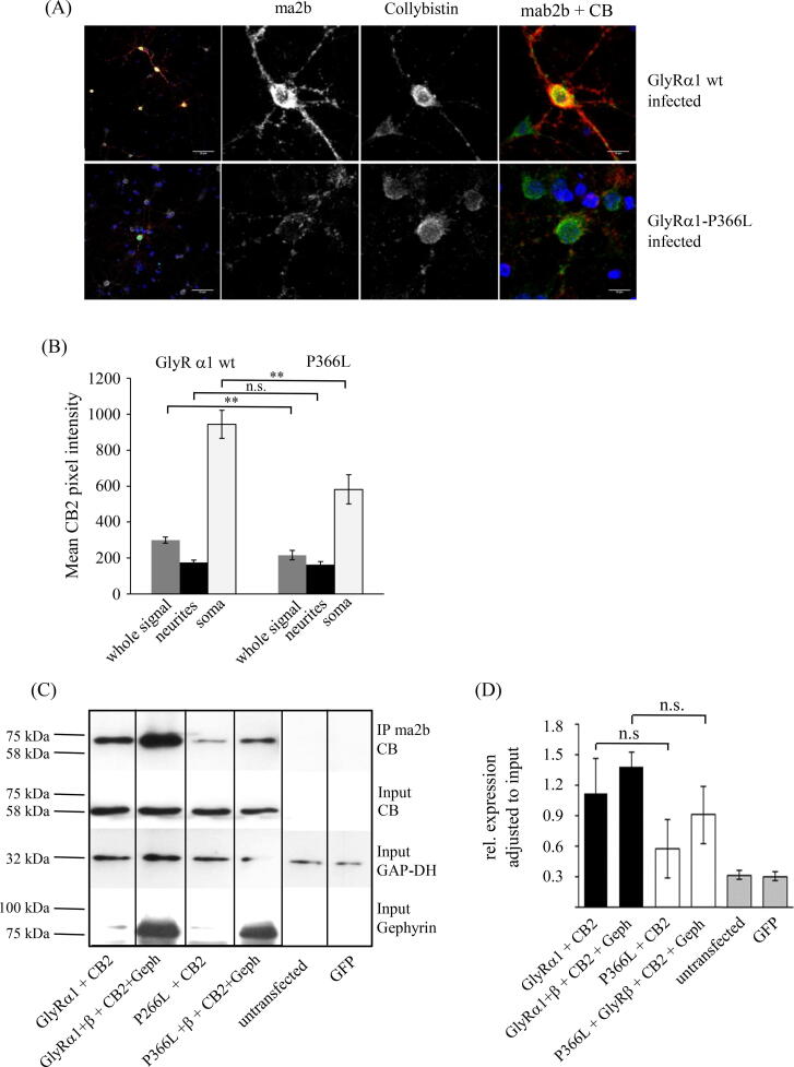
Results: Recombinantly expressed collybistin bound to isolated GlyRα1 TM3-4 loops in GST-pulldown assays. When the five proline residues P365A, P366A, P367A, P369A, P373A (GlyRα1P1-5A) located in the GlyRα1-PPII helix were replaced by alanines, the PPII secondary structure was disrupted. Recombinant GlyRα1P1-5A mutant subunits displayed normal cell surface expression and wildtype-like ion channel function, but binding to collybistin was abolished. The GlyRα1-collybistin interaction was independently confirmed by o-immunoprecipitation assays using full-length GlyRα1 subunits. Surprisingly, the interaction was not mediated by the SH3 domain of collybistin, but by its Pleckstrin homology (PH) domain. The mutation GlyRα1P366L, identified in a hyperekplexia patient, is also disrupting the PPII helix, and caused reduced collybistin binding.

Conclusion: Our data suggest a novel interaction between α1 GlyR subunits and collybistin, which is physiologically relevant in vitro and in vivo and may contribute to postsynaptic anchoring of glycine receptors.

Keywords: Collybistin; Gephyrin; Glycine receptor alpha1 subunit; Ion channel receptors; Pleckstrin homology domains; Polyproline II helix; Protein-protein interaction; SH3 domains; Synaptic anchoring.


Conflict of interest statement

The authors declare that they have no known competing financial interests or personal relationships that could have appeared to influence the work reported in this paper.

Figures

None
Graphical abstract
Fig. 1
Fig. 1
Localization and characterization of the GlyRα1 PPII helix. (A) Model of GlyRα1 with its predicted PPII helix in the TM3-4 loop (adapted from . (B) Sequence of wildtype GlyR in comparison with group mutant at position 365–373. (C) SDS-PAGE of GlyRα1 as well as GlyRα1P1-5A mutant TM3-4 loops after over-expression in E. coli cells and purification via Ni-NTA column and gel filtration chromatography. Lane 1: α1 wildtype TM3-4 loop; lane 2: GlyRα1P1-5A TM3-4 loop; lane 3: protein standard. Position of TM3-4 loop and 12 kDa is indicated. (D) CD spectra of α1-wt (solid line) and GlyRα1P1-5A (dashed line). All spectra were measured in 10 mM K-phosphate, pH 7.4 in a 1 cm cuvette at 22 °C. Eight single spectra were summed and the reference spectrum (10 mM K-phosphate, pH 7.4) was subtracted.
Fig. 2
Fig. 2
Expression and cellular distribution of GlyRα1 wildtype and GlyRα1P1-5A mutant subunits. (A) Western blot analysis of GlyRα1 subunits. 20 μg of membrane preparation was loaded per lane, primary antibody was mAb4a supernatant. Lane 1: α1 wildtype; lane 2: GlyRα1P1-5A; lane 3: protein standard; 48 kDa is indicated. (B) Immunofluorescence: HEK293 cells were transfected on cover slides and treated for immunocytochemistry with mAb4a and goat anti mouse Cy3. For surface expression no Triton X-100 was added. To detect the intracellular protein distribution, the cell membrane was permeabilized with Triton X-100. Controls: untransfected cells or GlyRα1 transfected cells treated with secondary antibody only are shown. Pictures were taken at 400× magnification. Scale bar indicates 10 µm.
Fig. 3
Fig. 3
Patch-clamp electrophysiological characterization of homomeric GlyRα1 wildtype and GlyRα1P1-5A mutant receptors. (A) Current responses of human wildtype GLRA1 (hs GlyRα1-wt) and mutant receptors GlyRα1P1-5A. Whole-cell patch clamp currents were recorded from transfected HEK293 cells at glycine concentrations between 10 and 1000 μM and a membrane potential of −50 mV. (B) Dose response curve of α1-wt (solid squares, solid line) and GlyRα1P1-5A (open circles and dashed line). EC50 values were 38.4 ± 3.4 μM for α1-wt (n = 20) and 56.9 ± 4.4 μM (n = 9) for the mutant receptor. Hill constants were 1.9 ± 0.1 and 2.0 ± 0.1 for wildtype and mutant, respectively. (C) Current potential curves were measured at 2 mM glycine in a potential range of −60 mV to +60 mV. Current responses were normalized to maximal currents at −60 mV. (D) Comparison of wildtype and mutant receptor dose responses; the difference was statistically significant (p = 0.016).
Fig. 4
Fig. 4
Interaction between collybistin and the GlyRα1 TM3-4 loop. (A) Schematic structure of collybistin variants 1–3. (B) Binding studies between α1-GlyR and collybistin I-II using GST pull down assay upon HEK293 cell transfection with CB1SH+-HA and CB2SH--HA. GST-fusion proteins of the isolated GlyRα1 wildtype or GlyRα1P1-5A TM3-4 loop were recombinantly expressed in E. coli BL21 cells, coupled to glutathione-sepharose beads and finally incubated with HEK293 cell lysates. Results are shown after Western Blot analysis. Lane 1: input, lane 2: GST control, lane 3: α1-wt TM3-4 loop, lane 4: GlyRα1P1-5A TM3-4 loop. C Co-immunoprecipitation of glycine receptors and collybistin splice variants. The GlyRα1-specific antibody mAb2b was used for co-precipitation. Lane 1: GlyRα1 + CB1 (1:1); lane 2: GlyRα1 + CB2 (1:1); lane 3: GlyRα1 + GlyRβ + CB2 + gephyrin (1:2:2:2); lane 4: untransfected cells; lane 5: GFP = mock control (left panel). Collybistin was detected at the appropriate molecular weight of 60 kDa (precipitated with GlyRα1 – see IP, upper panel and input control expression of collybistin - see input second panel; gephyrin at 93 kDa, and GAP-DH at 32 kDa. The observed shift in molecular weight between CB1 and CB2 is due to the presence of the SH3 domain in CB1 but not CB2. Right panel: Quantification of the relative collybistin expression normalized to GAPDH. At least 4 independent experiments have been performed and were used for quantification analysis. (D) Co-immunoprecipitation of GFP-CB2 or GFP-PH domain together with pCIS-HA-GlyRα1. Beads were coupled with mouse anti-HA antibody or mouse IgG, HEK293 cell lysates were incubated with antibody coupled beads (see methods). Samples were subjected to SDS PAGE and Western Blotting. Lane 1: input; lane 2: IgG control; lane 3: IP HA tag. Left panel: GFP-CB2; right panel: GFP-PH domain. (E) Electrophysiological data after co-expression of GlyRα1 with CB1 or CB2. Left panel: EC50 curve of GlyRα1 (solid squares, solid line); GlyRα1 co-expressed with CB1 (open circles, dashed line) and GlyRα1 co-expressed with CB2 (open triangle, dotted line). Right panel: comparison of GlyRα1 and co-transfection with CB1 and CB2. Differences were not significant (p > 0.05, one-way ANOVA).
Fig. 5
Fig. 5
Endogenous collybistin colocalizes with GlyRα1 in primary murine neurons. (A) Hippocampal neurons were infected with a lentivirus encoding either GlyRα1 wildtype or a pathological GlyRα1P366L variant carrying a mutation in the PPII helix. At DIV15, cells were co-stained for GlyRα1 (mAb2b, 1:500, red) and endogenous collybistin (polyclonal rabbit anti-collybistin antibody, 1:500, green). Note, infected cells were controlled by bidirectional GFP expression. Hence, collybistin was stained with the secondary goat-anti-rabbit Cy5 antibody and is shown in false color. Nuclear DAPI staining is shown in blue. White bar in left overview panels refers to 50 µm, white bar in zoomed pictures (second to fourth lane) refers to 10 µm. (B) Quantification of the collybistin intensities determined in mAb2b (GlyRα1) clusters. Mean intensities are shown for whole cells, neurites and soma. P-values to represent level of significance are indicated **p < 0.01, n.s. = non-significant. (C) Co-immunoprecipitation of the GlyRα1 wildtype and the pathological GlyRα1P366L variant with collybistin following overexpression in HEK293 cells. Both variants were expressed with CB2 (ratio 1:1) only to detect direct interaction and co-transfected with the GlyRβ subunit and gephyrin (ratio 1:2:2:2) to detect if these structural GlyR complex proteins promote interaction with collybistin. Precipitated collybistin is shown at the appropriate molecular weight of 60 kDa in the upper panel, collybistin input second panel, GAP-DH (32 kDa) input control third panel, gephyrin (93 kDa) expression lower panel. Note, although the collybistin expression is similar (input), collybistin precipitated more efficiently in the presence of GlyRβ and gephyrin (abbreviation Geph). (D) Quantification of relative collybistin expression in the presence of collybistin only or together with GlyRβ and gephyrin. Four independent experiments have been performed and were used for analysis; n.s. non-significant.

Similar articles

Cited by

References

    1. Breitinger HG. Glycine Receptors. Chichester: eLS. John Wiley & Sons Ltd; 2014.
    1. Lynch J.W. Native glycine receptor subtypes and their physiological roles. Neuropharmacology. 2009;56:303–309. - PubMed
    1. Grunewald N., Jan A., Salvatico C., Kress V., Renner M., Triller A. Sequences flanking the gephyrin-binding site of GlyRbeta tune receptor stabilization at synapses. eNeuro. 2018;5 - PMC - PubMed
    1. Kneussel M., Betz H. Receptors, gephyrin and gephyrin-associated proteins: novel insights into the assembly of inhibitory postsynaptic membrane specializations. J Physiol. 2000;525(Pt 1):1–9. - PMC - PubMed
    1. Ogino K., Yamada K., Nishioka T., Oda Y., Kaibuchi K., Hirata H. Phosphorylation of Gephyrin in Zebrafish Mauthner cells governs glycine receptor clustering and behavioral desensitization to sound. J Neurosci. 2019;39:8988–8997. - PMC - PubMed

LinkOut - more resources