

Finite Element Modelling and Experimental Validation of the Enamel Demineralisation Process at the Rod Level
- PMID: 33842014
- PMCID: PMC8020348
- DOI: 10.1016/j.jare.2020.08.018
Finite Element Modelling and Experimental Validation of the Enamel Demineralisation Process at the Rod Level
Abstract
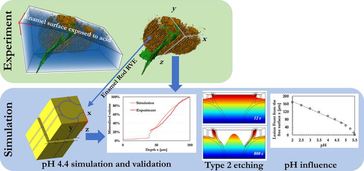
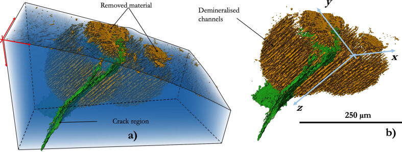
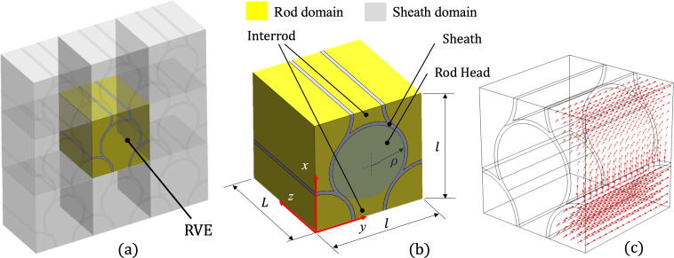
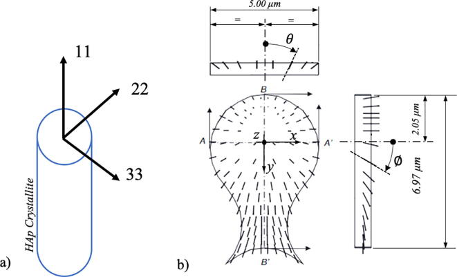
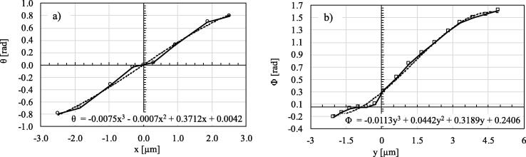
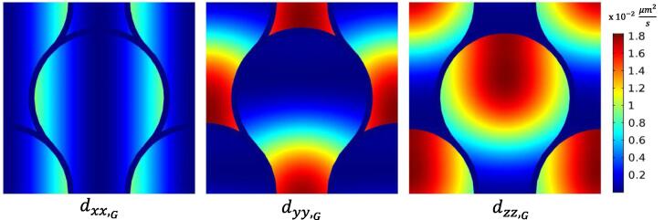
In the past years, a significant amount of effort has been directed at the observation and characterisation of caries using experimental techniques. Nevertheless, relatively little progress has been made in numerical modelling of the underlying demineralisation process. The present study is the first attempt to provide a simplified calculation framework for the numerical simulation of the demineralisation process at the length scale of enamel rods and its validation by comparing the data with statistical analysis of experimental results. FEM model was employed to simulate a time-dependent reaction-diffusion equation process in which H ions diffuse and cause demineralisation of the enamel. The local orientation of the hydroxyapatite crystals was taken into account. Experimental analysis of the demineralising front was performed using advanced high-resolution synchrotron X-ray micro-Computed Tomography. Further experimental investigations were conducted by means of SEM and STEM imaging techniques. Besides establishing and validating the new modelling framework, insights into the role of the etchant solution pH level were obtained. Additionally, some light was shed on the origin of different types of etching patterns by simulating the demineralisation process at different etching angles of attack. The implications of this study pave the way for simulations of enamel demineralisation within different complex scenarios and across the range of length scales. Indeed, the framework proposed can incorporate the presence of chemical species other than H ions and their diffusion and reaction leading to dissolution and re-precipitation of hydroxyapatite. It is the authors' hope and aspiration that ultimately this work will help identify new ways of controlling and preventing caries.
Keywords: Demineralisation simulation; Dental demineralisation; Enamel; FEM; Reaction-diffusion; Synchrotron CT.
© 2021 The Authors. Published by Elsevier B.V. on behalf of Cairo University.
Conflict of interest statement
The authors declare that they have no known competing financial interests or personal relationships that could have appeared to influence the work reported in this paper.
Figures
References
-
- N. Pitts, Understanding dental caries - From pathogenesis to prevention and therapy, Understanding Dental Caries: From Pathogenesis to Prevention and Therapy, Springer International Publishing, 2016, pp. 3–9.
-
- Schüpbach P., Guggenheim B., Lutz F. Human root caries: histopathology of initial lesions in cementum and dentin. J Oral Pathol Med. 1989;18(3):146–156. - PubMed
-
- Ash M.M. W.B. Saunders; 1993. Wheeler's dental anatomy, physiology, and occlusion.
-
- Chandra Satish, Chandra Shaleen, Chandra MIthilesh, Chandra Grish, Chandra N. Textbook of dental and oral histology with embryology and multiple choice questions. Jaypee Brothers Medical Publishers; 2010.
-
- Chiego D, Chiedo DJ. Essentials of oral histology and embryology. Elsevier; 2013.
LinkOut - more resources
Full Text Sources
Other Literature Sources
Miscellaneous
