Review
. 2021 Apr;11(2):e48-e63.
doi: 10.1212/CPJ.0000000000000908.

Neurology and the COVID-19 Pandemic: Gathering Data for an Informed Response

Affiliations
Review

Neurology and the COVID-19 Pandemic: Gathering Data for an Informed Response

Brigit High et al. Neurol Clin Pract. 2021 Apr.

Abstract

Purpose of review: The current coronavirus disease 2019 (COVID-19) pandemic caused by the severe acute respiratory syndrome coronavirus 2 (SARS-CoV-2) is one of the greatest medical crises faced by our current generation of health care providers. Although much remains to be learned about the pathophysiology of SARS-CoV-2, there is both historical precedent from other coronaviruses and a growing number of case reports and series that point to neurologic consequences of COVID-19.

Recent findings: Olfactory/taste disturbances and increased risk of strokes and encephalopathies have emerged as potential consequences of COVID-19 infection. Evidence regarding whether these sequelae result indirectly from systemic infection or directly from neuroinvasion by SARS-CoV-2 is emerging.

Summary: This review summarizes the current understanding of SARS-CoV-2 placed in context with our knowledge of other human coronaviruses. Evidence and data regarding neurologic sequelae of COVID-19 and the neuroinvasive potential of human coronaviruses are provided along with a summary of patient registries of interest to the Neurology community.


Figures

Figure 1
Figure 1. Schematic of a SARS-CoV-2 Virion
The image depicts the severe acute respiratory syndrome coronavirus 2 (SARS-CoV-2) virion with lipid bilayer membrane and shows the structural spike (S), membrane (M), envelope (E), and nucleocapsid (N) proteins. The virion contains a single-strand, positive-sense RNA (+ssRNA) genome surrounded by its N protein chaperone. The S, M, E, and N proteins are possible targets for vaccine development.
Figure 2
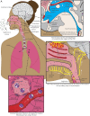
Figure 2. Proposed Pathways of SARS-CoV-2 Systemic Infection and Neuroinvasion
(A) Initial infection by severe acute respiratory syndrome coronavirus 2 (SARS-CoV-2) with viral entry into the nasopharynx, oropharynx, larynx, and lungs. (B) Magnified cross-section of a lung alveolus and capillary. SARS-CoV-2 is known to infect type II pneumocytes as well as endothelial cells, which express the angiotensin-converting enzyme 2 (ACE-2). From the lungs, the virus could enter the vasculature and circulate to other organs, resulting in multiorgan infection and sepsis. Inflammation of cells such as neutrophils, monocytes, and lymphocytes could contribute to viral access to the vasculature due capillary leakage and direct tissue damage. This may also be an initial site for viral infection of immune cells such as macrophages and lymphocytes, which have been shown to become infected by other coronaviruses, such as SARS-CoV-1, and express the ACE-2 receptor. (C) Sagittal cross-section of the brain at the level of the third and fourth ventricles with surrounding structures. Hematogenous spread could lead to direct viral infection of the CNS or infection of the CNS via the circumventricular organs (CVOs) including the subfornical organs, the vascular organ of the lamina terminalis (OVLT), the median eminence, and the area postrema, which do lack a blood-brain barrier and have been shown to express ACE-2. SARS-CoV-2 may also access the CNS via the choroid plexus, depicted on the roof of the third ventricle and in the fourth ventricle. Virus could infect the vasculature of these regions, which expresses ACE-2 or traffic into the CNS via infected lymphocytes. (D) Sagittal cross-section of the upper respiratory tract. SARS-CoV-2 infection has been associated with hyposmia and hypogeusia in a significant number of patients. ACE-2 has been found in the non-neuronal cells of the olfactory system, suggesting dysfunction damage to non-neuronal cells as a cause of anosmia. Hypogeusia could be caused by viral infection of gustatory neurons of cranial nerves VII (chorda tympani of the facial nerve), CN IX, and CN X, all of which project to the nucleus of the solitary tract in the brainstem and are known to express ACE-2. Gustatory pathways could contribute to viral CNS access to the brainstem that ultimately contributes to respiratory dysfunction. Infection of the brainstem could worsen respiratory distress in patients with severe COVID-19. For all panels, SARS-CoV-2 virions are depicted as green dots. Black or white arrows show possible directions of viral spread in each organ or tissue.

References

    1. Perlman S, Netland J. Coronaviruses post-SARS: update on replication and pathogenesis. Nat Rev Microbiol 2009;7:439–450. - PMC - PubMed
    1. Coronaviridae Study Group of the International Committee on Taxonomy of Viruses. The species Severe acute respiratory syndrome-related coronavirus: classifying 2019-nCoV and naming it SARS-CoV-2. Nat Microbiol 2020;5:536–544. - PMC - PubMed
    1. Desforges M, Le Coupanec A, Dubeau P, et al. Human coronaviruses and other respiratory viruses: underestimated opportunistic pathogens of the central nervous system? Viruses 2019;12:14. - PMC - PubMed
    1. Li F. Receptor recognition mechanisms of coronaviruses: a decade of structural studies. J Virol 2015;89:1954–1964. - PMC - PubMed
    1. Tang X, Wu C, Li X, et al. On the origin and continuing evolution of SARS-CoV-2. Natl Sci Rev 2020;7:1012–1023. - PMC - PubMed