

Rapidly Growing Huge Lower Back Malignant Peripheral Nerve Sheath Tumour
- PMID: 33842103
- PMCID: PMC8020617
- DOI: 10.7759/cureus.13712
Rapidly Growing Huge Lower Back Malignant Peripheral Nerve Sheath Tumour
Abstract
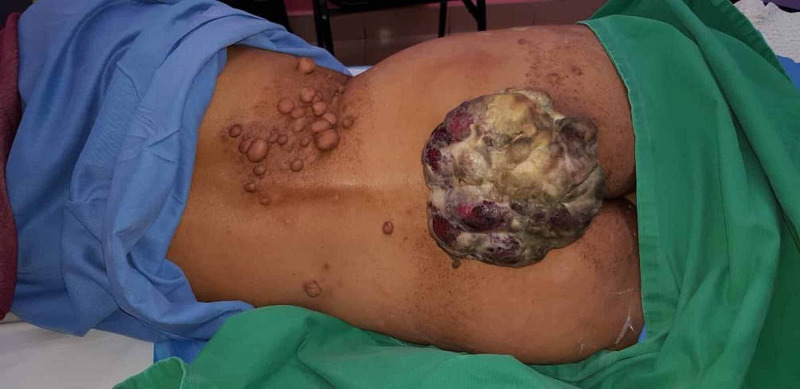
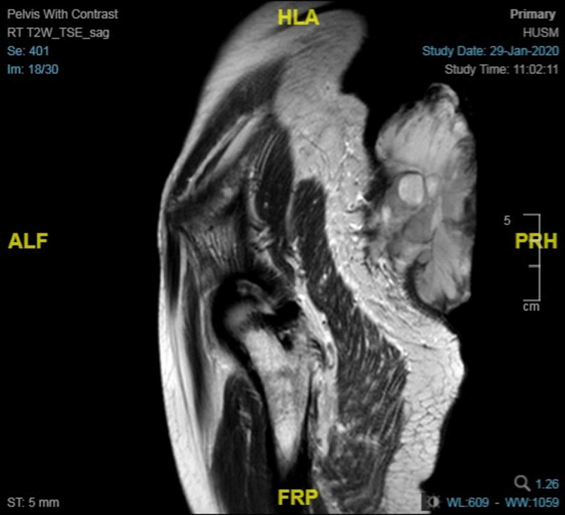
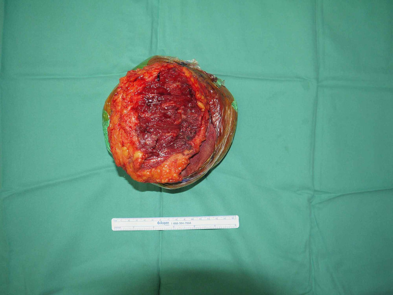
Malignant peripheral nerve sheath tumours (also called neurofibrosarcomas) are a rare, highly aggressive soft tissue sarcomas that arise from the peripheral nerves or cells associated with the nerve sheath, such as Schwann cells, peri-neural cells and fibroblasts. It is representing 10% of all soft tissue sarcomas in which it is considered as an extremely rare malignancy, especially in patients with neurofibromatosis type I. In the general population, it affects approximately 1 in 100,000 people. This article is reporting a 56-year-old Malay female patient who is a known case of neurofibromatosis type I for 20 years, presented with a lower back, pruritic, gradually increasing swelling during the last five months. Last month before the presentation, the lesion rapidly grows, reaching a size of (15×15 cm), accompanied by foul-smelling discharge and pain exacerbated with movement. Although no history of preceding trauma or accident, the mass bleeds within contact. In conclusion, only a few cases of giant malignant peripheral nerve sheath tumours reported in the literature describing its location and growth progression. We present a massive, extremely rapid growth of cutaneous exophytic malignant peripheral nerve sheath tumours over the lower back.
Keywords: malignant peripheral nerve sheath tumour; neurofibromatosis; neurofibrosarcomas.
Copyright © 2021, Abuzarifa et al.
Conflict of interest statement
The authors have declared that no competing interests exist.
Figures
Similar articles
-
Malignant Peripheral Nerve Sheath Tumour Presenting as Sciatica in a Patient with Neurofibromatosis Type 1: Is Magnetic Resonance Imaging an Effective Screening Tool?Eur J Case Rep Intern Med. 2024 Oct 24;11(11):004818. doi: 10.12890/2024_004818. eCollection 2024. Eur J Case Rep Intern Med. 2024. PMID: 39525431 Free PMC article.
-
Malignant peripheral nerve cell tumour.J Maxillofac Oral Surg. 2010 Mar;9(1):68-71. doi: 10.1007/s12663-010-0019-6. Epub 2010 Jun 4. J Maxillofac Oral Surg. 2010. PMID: 23139572 Free PMC article.
-
Malignant peripheral nerve sheath tumour presenting as a pneumothorax.Br J Radiol. 2011 Oct;84(1006):e197-9. doi: 10.1259/bjr/27394681. Br J Radiol. 2011. PMID: 21933975 Free PMC article.
-
[Malignant intracerebral nerve sheath tumours: Two case reports and complete review of the literature cases].Cancer Radiother. 2016 Apr;20(2):119-32. doi: 10.1016/j.canrad.2015.07.157. Epub 2016 Feb 27. Cancer Radiother. 2016. PMID: 26934901 Review. French.
-
Fungating malignant peripheral nerve sheath tumor arising from a slow-growing mass in the forearm: a case report and review of the literature.J Med Case Rep. 2020 Jul 7;14(1):91. doi: 10.1186/s13256-020-02427-4. J Med Case Rep. 2020. PMID: 32631436 Free PMC article. Review.
References
-
- Giant malignant peripheral nerve sheath tumor of the scalp. Fukushima S, Kageshita T, Wakasugi S, et al. J Dermatol. 2006;33:865–868. - PubMed
-
- Malignant peripheral nerve sheath tumors of the buttock and lower extremity. A study of 43 cases. Hruban RH, Shiu MH, Senie RT, et al. Cancer. 1990;66:1253–1265. - PubMed
Publication types
LinkOut - more resources
Full Text Sources
Other Literature Sources
Research Materials