Case Reports
. 2021 Mar 16:37:101635.
doi: 10.1016/j.eucr.2021.101635. eCollection 2021 Jul.

Penetrating scrotal injury in childhood

Affiliations
Case Reports

Penetrating scrotal injury in childhood

Muhammad Asykar Palinrungi et al. Urol Case Rep. .

Abstract

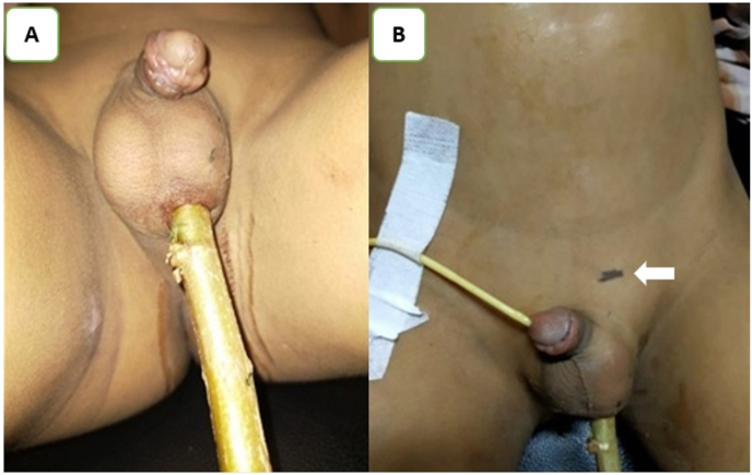
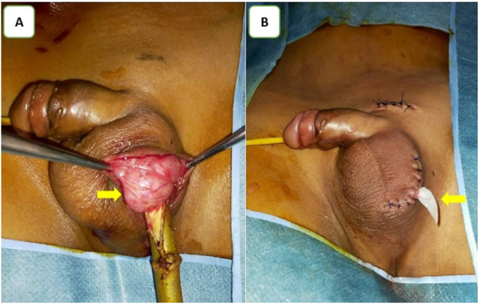
An 11-year-old boy was referred to the emergency room (ER) because of a stab wound on the scrotum due to a tree trunk. The external genitalia computed tomography (CT) scan revealed a 5-cm long stick that was stuck in his left hemiscrotum; however, the testis was normal. Scrotal exploration and debridement of non-viable tissue was performed, and no testicular or surrounding structure injury was found. At the postoperative evaluation 1 month later, the surgical wound and the patient's general condition were satisfactory.

Keywords: Foreign body; Genitalia; Penetrating injury; Scrotal trauma.


Conflict of interest statement

The authors declare that they have no conflict of interests.

Figures

Fig. 1
Fig. 1
Image of the patient's stab wound to the scrotum before surgery. 1A. A stick with an entry site in the left hemiscrotum. 1B. The tip of the stick was at the upper level of the left pubic bone (white arrow).
Fig. 2
Fig. 2
Non-contrast CT and a three-dimensional CT imaging before the surgery. 2A and 2B. A 5-cm stick (blue arrow) was stuck in the left hemiscrotum. 2C. The foreign body (blue arrow) and the left testis (yellow arrow), which was normal, are shown using non-contrast CT. 2D. A three-dimensional CT of the patient's external genitalia showed the foreign body (blue arrow). (For interpretation of the references to colour in this figure legend, the reader is referred to the Web version of this article.)
Fig. 3
Fig. 3
Intraoperative image. 3A. The testis (arrow) and spermatic cord was normal upon scrotal exploration and debridement. 3B. A Penrose drain (arrow) was left on the side of the wound.

References

    1. Ferlise V.J., Haranto V.H., Ankem M.K., Barone J.G. Management of penetrating scrotal injury. Pediatr Emerg Care. 2002;18 https://journals.lww.com/pec-online/Fulltext/2002/04000/Management_of_pe... - PubMed
    1. Randhawa H., Blankstein U., Davies T. Scrotal trauma: a case report and review of the literature. Can Urol Assoc J. 2019;13 doi: 10.5489/cuaj.5981. - DOI - PMC - PubMed
    1. Owusu Ofori E., Bin Alhassan B.A., Essoun S., Asante-Asamani A., Maison P. Penetrating scrotal injury: two unusual case reports in children and brief review of literature. J Adv Med Med Res. 2020:39–44. doi: 10.9734/jammr/2020/v32i930480. - DOI
    1. Lynch T., Martinez-Pineiro L., Plas E. EAU guidelines on urological trauma. Eur Urol. 2005;47:1–15. doi: 10.1016/j.eururo.2004.07.028. - DOI - PubMed
    1. Morey A.F., Metro M.J., Carney K.J., Miller K.S., McAninch J.W. Consensus on genitourinary trauma: external genitalia. BJU Int. 2004;94:507–515. doi: 10.1111/j.1464-410X.2004.04993.x. - DOI - PubMed

Publication types

LinkOut - more resources