. 2021 Mar 17:8:599-606.
doi: 10.1016/j.toxrep.2021.03.011. eCollection 2021.

Combination therapy of N-acetyl-L-cysteine and S-2(2-aminoethylamino) ethylphenyl sulfide for sulfur mustard induced oxidative stress in mice

Affiliations

Combination therapy of N-acetyl-L-cysteine and S-2(2-aminoethylamino) ethylphenyl sulfide for sulfur mustard induced oxidative stress in mice

Alka Gupta et al. Toxicol Rep. .

Abstract

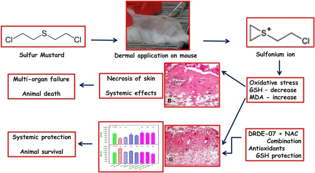
Introduction: Sulfur mustard (SM) is chemically, bis(2-chloroethyl) sulfide and a strong alkylating agent that causes cytotoxicity and blisters on skin. In laboratory animal models, SM is extremely lethal. Since no specific antidote has been proposed, decontamination upon contact is the recommended procedure. Several antidotes have been screened for SM, and in that sulfanyl compounds, N-acetyl-l-cysteine (NAC) and S-2(2-aminoethylamino) ethylphenyl sulfide (DRDE-07) showed good protection. Since they showed protection at high doses, the aim of this study was to evaluate the efficacy in combination at low dose, for percutaneously administered SM in mice.

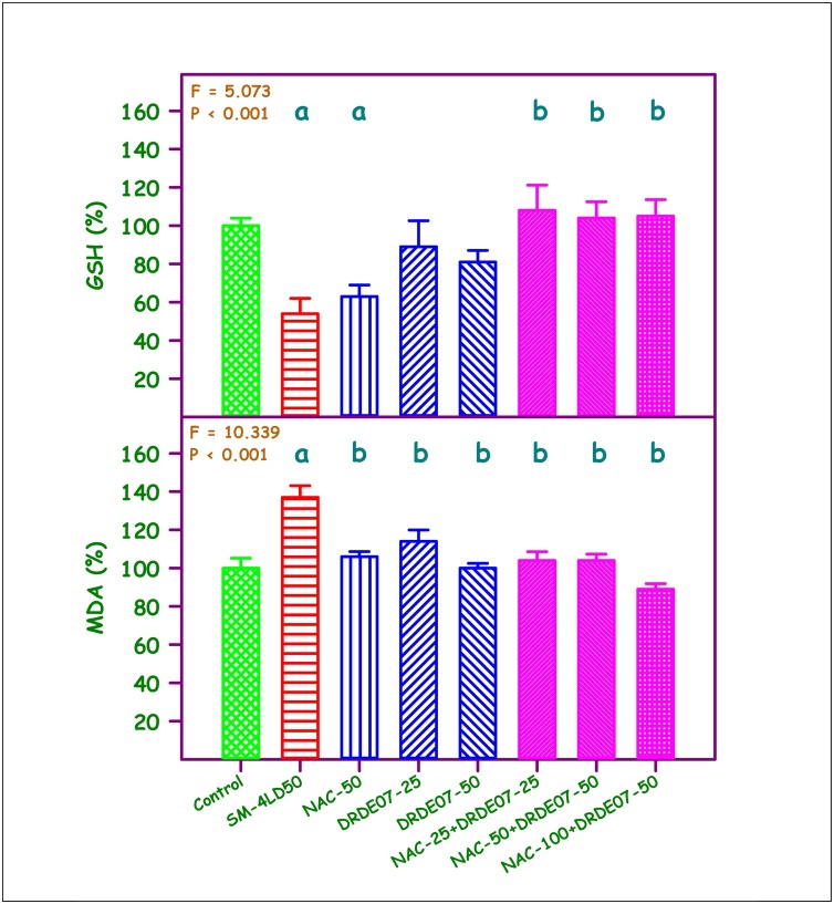
Material and methods: 4 LD50 of SM (32.4 mg/kg) was administered, and NAC (50 mg/kg), DRDE-07 (25 and 50 mg/kg) and their combinations were evaluated as 30 min pre-treatment by single oral administration.

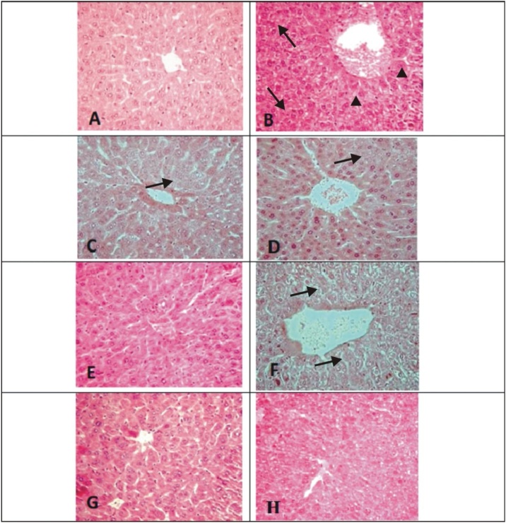
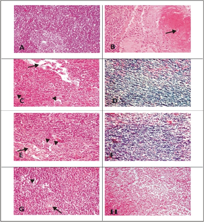
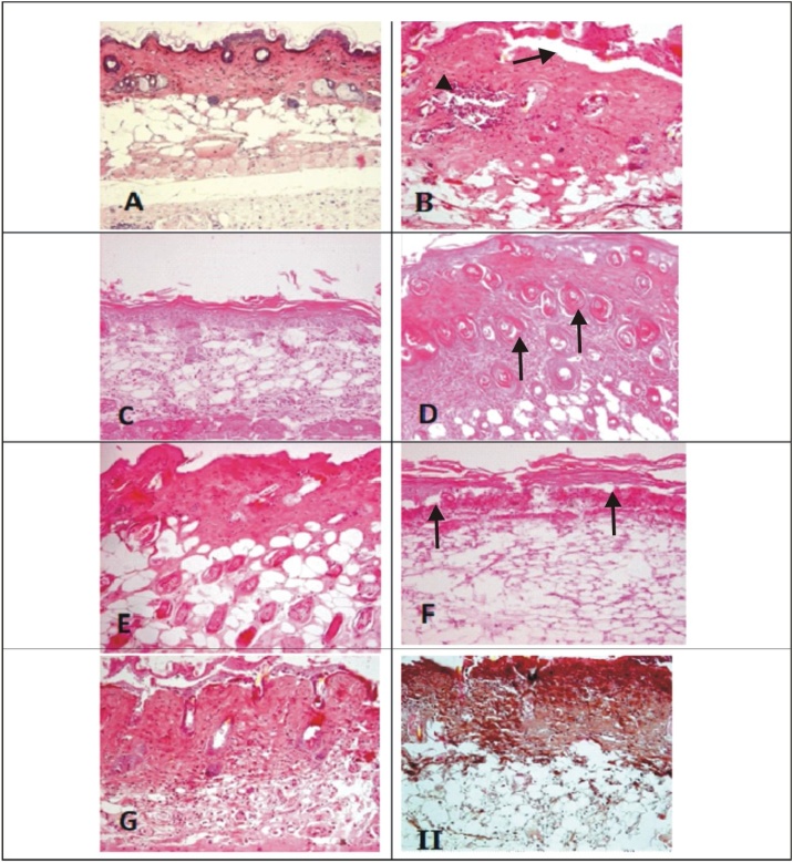
Result: After 72 h of SM exposure, significant decrease in body weight, decrease in hepatic reduced glutathione, and increase in hepatic malondialdehyde were observed (P < 0.001), showing oxidative stress. The combination of NAC (100 mg/kg) and DRDE-07 (50 mg/kg) showed significant protection (P < 0.01). The severe histopathological lesions induced by SM in liver, spleen and skin were also considerably reduced by the combination.

Conclusion: The combination of NAC and DRDE-07 having sulfanyl groups, will be promising antioxidants and an effective antidote for SM toxicity.

Keywords: Antidote; DRDE-07; Glutathione; Malondialdehyde; Mice; N-acetylcysteine; Oxidative stress; Sulfur mustard.


Conflict of interest statement

The authors declare that they have no known competing financial interests or personal relationships that could have appeared to influence the work reported in this paper.

Figures

None
Graphical abstract
Fig. 1
Fig. 1
Percent change in reduced glutathione (GSH) and malondialdehyde (MDA) levels in liver. Control values - GSH = 2.79 + 0.12 μmol/g tissue, MDA = 1.17 + 0.17 nmol/g tissue. Values are mean + SEM (n = 6 each). aSignificantly different from control group. bSignificantly different from SM group.
Fig. 2
Fig. 2
Photomicrograph of liver tissue (H & E; 100x). Representative image of each group (A) Control mice showing normal hepatic chord, hepatocytes, central canal and kupffer cells; (B) SM-4LD50 arrow showing severe necrosis, arrowheads showing ballooning of hepatocytes and distortion of lobular pattern; (C) NAC-50 arrow showing reduction of severity of lesions as compared to B; (D) DRDE07−25 arrow showed slight decrease in severity of degenerative changes than B; (E) DRDE07−50; (F) NAC-25+DRDE07−25 arrow showing minimal degenerative changes as compared to B; (G) NAC-50+DRDE07−50; (H) NAC-100+DRDE07−50
Fig. 3
Fig. 3
Photomicrograph of spleen tissue (H & E;100x). Representative image of each group (A) Control mice spleen showing normal spleenic histology with germinal center, red pulp and marginal zone of white pulp; (B) SM-4LD50 arrow showing severe necrosis/apoptosis; (C) NAC-50 arrow showing fibrinous exudates and arrowheads showing necrosis/apoptosis; (D) DRDE07−25; (E) DRDE07−50 arrow showing fibrinous exudates and arrowheads showing necrosis/apoptosis; (F) NAC-25+DRDE07−25 showing lymphoid depletion; (G) NAC-50+DRDE07−50 arrow showing fibrinous and arrowheads showing lymphoid depletion ; (H) NAC-100+DRDE07−50
Fig. 4
Fig. 4
Photomicrograph of skin (H & E; 100x). Representative image of each group (A) Control mice skin section showing normal arrangement of epidermis, dermis, hair follicles and sebaceous glands; (B) SM-4LD50 arrow showing inflammation, atrophy dermoepidermal sepration; (C) NAC-50 showing hyalinization; (D) DRDE07−25; (E) DRDE07−50 (F) NAC-25+DRDE07−25 arrow showing coagulative necrosis of epidermis penetrating deep into dermis along with necrotic inflammatory cells; (G) NAC-50+DRDE07−50 showing mild to moderate decrease in severity of skin lesions compared to B; (H) NAC-100+DRDE07−50.

Similar articles

Cited by

References

    1. Romano J.A., Lukey B.J., Salem H. CRC Press; Boca Raton FL. USA: 2008. Chemical Warfare Agents: Chemistry, Pharmacology, Toxicology, and Therapeutics.
    1. Kehe K., Szinicz L. Medical aspects of sulphur mustard poisoning. Toxicology. 2005;214:198–209. doi: 10.1016/j.tox.2005.06.014. - DOI - PubMed
    1. Saladi R.N., Smith E., Persaud A.N. Mustard: a potential agent of chemical warfare and terrorism. Clin. Exp. Dermatol. 2006;31:1–5. doi: 10.1111/j.1365-2230.2005.01945.x. - DOI - PubMed
    1. Graham J.S., Chilcott R.P., Rice P., Milner S.M., Hurst C.G., Maliner B.I. Wound healing of cutaneous sulfur mustard injuries: strategies for the development of improved therapies. J. Burns Wounds. 2005;4 e1. Published 2005 Jan 5. - PMC - PubMed
    1. Vijayaraghavan R., Kulkarni A., Pant S.C., Kumar P., Rao P.V., Gupta N. Differential toxicity of sulfur mustard administered through percutaneous, subcutaneous, and oral routes. Toxicol. Appl. Pharmacol. 2005;202:180–188. doi: 10.1016/j.taap.2004.06.020. - DOI - PubMed