. 2021 Mar 26:11:610338.
doi: 10.3389/fonc.2021.610338. eCollection 2021.

Machine Learning-Based Radiomics Nomogram for Detecting Extramural Venous Invasion in Rectal Cancer

Affiliations

Machine Learning-Based Radiomics Nomogram for Detecting Extramural Venous Invasion in Rectal Cancer

Siye Liu et al. Front Oncol. .

Abstract

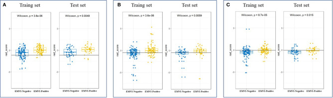
Objective: To establish and validate a radiomics nomogram based on the features of the primary tumor for predicting preoperative pathological extramural venous invasion (EMVI) in rectal cancer using machine learning.

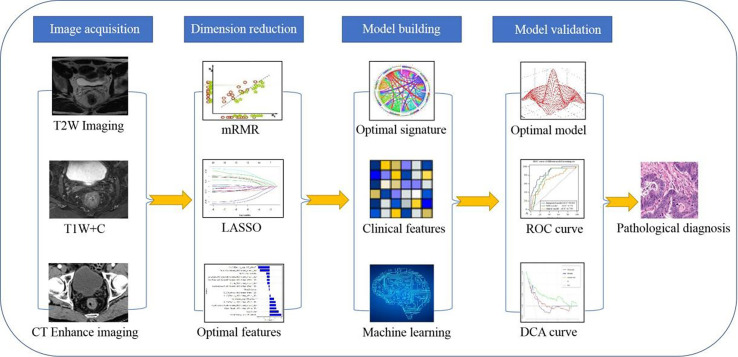
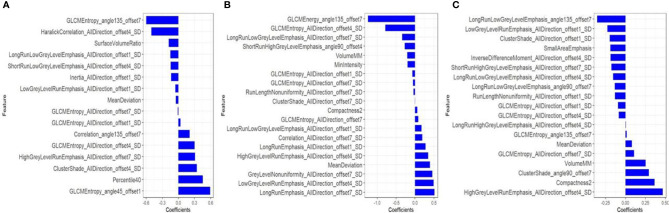
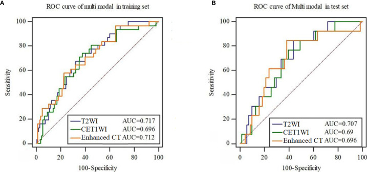
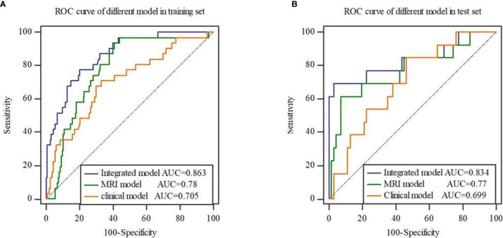
Methods: The clinical and imaging data of 281 patients with primary rectal cancer from April 2012 to May 2018 were retrospectively analyzed. All the patients were divided into a training set (n = 198) and a test set (n = 83) respectively. The radiomics features of the primary tumor were extracted from the enhanced computed tomography (CT), the T2-weighted imaging (T2WI) and the gadolinium contrast-enhanced T1-weighted imaging (CE-TIWI) of each patient. One optimal radiomics signature extracted from each modal image was generated by receiver operating characteristic (ROC) curve analysis after dimensionality reduction. Three kinds of models were constructed based on training set, including the clinical model (the optimal radiomics signature combining with the clinical features), the magnetic resonance imaging model (the optimal radiomics signature combining with the mrEMVI status) and the integrated model (the optimal radiomics signature combining with both the clinical features and the mrEMVI status). Finally, the optimal model was selected to create a radiomics nomogram. The performance of the nomogram to evaluate clinical efficacy was verified by ROC curves and decision curve analysis curves.

Results: The radiomics signature constructed based on T2WI showed the best performance, with an AUC value of 0.717, a sensitivity of 0.742 and a specificity of 0.621. The radiomics nomogram had the highest prediction efficiency, of which the AUC was 0.863, the sensitivity was 0.774 and the specificity was 0.801.

Conclusion: The radiomics nomogram had the highest efficiency in predicting EMVI. This may help patients choose the best treatment strategy and may strengthen personalized treatment methods to further optimize the treatment effect.

Keywords: computed tomography; extramural venous invasion; magnetic resonance imaging; prediction; radiomics; rectal cancer.


Conflict of interest statement

The authors declare that the research was conducted in the absence of any commercial or financial relationships that could be construed as a potential conflict of interest.

Figures

Figure 1
Figure 1
Research flow chart of the radiomics model.
Figure 2
Figure 2
After dimensionality reduction by mRMR and LASSO, 16, 20 and 19 radiomics features were finally selected from CT-enhanced images (A), T2WI (B) and CE-T1WI (C) to construct a radiomics signature. The blue bar indicates the weight value of the radiomics features.
Figure 3
Figure 3
ROC curves of the radiomics signature constructed by each mode in the training set (A) and test set (B).
Figure 4
Figure 4
Scatter plot between EMVI-negative (blue dots) and EMVI-positive (yellow dots) rad scores calculated by radiomics signatures constructed by T2WI (A), CE-T1WI (B) and CT-enhanced images (C) in the training and test sets.
Figure 5
Figure 5
ROC curves of three models in the training set (A) and test set (B). The results show that the integrated model has the highest AUC value.
Figure 6
Figure 6
Radiomics nomogram for detecting EMVI (A). In the nomogram, a vertical line was drawn according to the value of the rad score to determine the corresponding value of points. The points of mrEMVI and differentiation stage can also be determined in the same way. The total points were the sum of the three points above. Finally, a vertical line was drawn according to the value of the total points to determine the probability of EMVI. The calibration curve of the radiomics nomogram for EMVI in the training set (B) and test set (C). A dashed line indicated the reference line where an ideal nomogram would lie. A dotted line indicated the performance of the nomogram, while the solid line indicated bias correction in the nomogram. DCA curve (D) for the integrated model, MRI model and clinical model predicting EMVI in the dataset. The graphs showed that the integrated model had the greatest net benefit. The risk classification performance of the integrated model in the training and test set (E). *P < 0.05.

Similar articles

Cited by

References

    1. Stewart DB, Dietz DW. Total mesorectal excision: what are we doing? Clin Colon Rectal Surg (2007) 20(3):190–202. 10.1055/s-2007-984863 - DOI - PMC - PubMed
    1. Nougaret S, Reinhold C, Mikhael HW, Rouanet P, Bibeau F, Brown G. The use of MR imaging in treatment planning for patients with rectal carcinoma: have you checked the “DISTANCE”? Radiology (2013) 268(2):330–44. 10.1148/radiol.13121361 - DOI - PubMed
    1. Liu L, Liu M, Yang Z, He W, Wang Z, Jin E. Correlation of MRI-detected extramural vascular invasion with regional lymph node metastasis in rectal cancer. Clin Imag (2016) 40(3):456–60. 10.1016/j.clinimag.2016.01.007 - DOI - PubMed
    1. McClelland D, Murray GI. A Comprehensive Study of Extramural Venous Invasion in Colorectal Cancer. PLoS One (2015) 10(12):e0144987. 10.1371/journal.pone.0144987 - DOI - PMC - PubMed
    1. Messenger DE, Driman DK, Kirsch R. Developments in the assessment of venous invasion in colorectal cancer: implications for future practice and patient outcome. Hum Pathol (2012) 43(7):965–73. 10.1016/j.humpath.2011.11.015 - DOI - PubMed

LinkOut - more resources