

Case Report: Cutaneous Squamous Cell Carcinoma Arising From the Ulcer of the Lesions of Dupuytren's Disease on the Palm
- PMID: 33842344
- PMCID: PMC8027102
- DOI: 10.3389/fonc.2021.638395
Case Report: Cutaneous Squamous Cell Carcinoma Arising From the Ulcer of the Lesions of Dupuytren's Disease on the Palm
Abstract
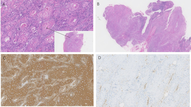
Dupuytren's disease is a benign fibromatosis that mainly involves the fascia of the palm and digits. The relationship between Dupuytren's disease and the evolution of cutaneous squamous cell carcinoma is still unclear. Here we report the case of a 52-year-old female with squamous cell carcinoma arising from the ulcer of the lesions of Dupuytren's disease on the left palm. To our knowledge, this is the first reported case in the English literature of squamous cell carcinoma on the palm of someone with Dupuytren's disease.
Keywords: Dupuytren’s disease; palm; skin cancer; squamous cell carcinoma; surgical excision.
Copyright © 2021 Sun, Fu, Li, Fang and Qiao.
Conflict of interest statement
The authors declare that the research was conducted in the absence of any commercial or financial relationships that could be construed as a potential conflict of interest.
Figures


Similar articles
-
[Squamous cell carcinoma in Dupuytren's disease--a case report].Handchir Mikrochir Plast Chir. 2011 Feb;43(1):54-6. doi: 10.1055/s-0030-1268480. Epub 2011 Jan 11. Handchir Mikrochir Plast Chir. 2011. PMID: 21225571 German.
-
Dupuytren's disease of the wrist.Hand Surg. 2001 Dec;6(2):235-7. doi: 10.1142/s0218810401000734. Hand Surg. 2001. PMID: 11901473
-
Dupuytren's contracture following burn injury of the hand: A case report and review of literature.Can J Plast Surg. 2008 Spring;16(1):49-51. doi: 10.1177/229255030801600105. Can J Plast Surg. 2008. PMID: 19554166 Free PMC article.
-
A study of the repeatability of the diagnosis of Dupuytren's contracture and its prevalence in the grampian region.J Hand Surg Br. 1993 Apr;18(2):258-61. doi: 10.1016/0266-7681(93)90124-x. J Hand Surg Br. 1993. PMID: 8501390 Review.
-
[A coexistence of the Dupuytren's disease and malignant neoplasms: a review].Ann Acad Med Stetin. 2013;59(1):15-7. Ann Acad Med Stetin. 2013. PMID: 24734329 Review. Polish.
Cited by
-
Giant Primary Cutaneous Myoepithelial Carcinoma of the Left Thigh With Inguinal and Pelvic Lymph Node Metastases.Cureus. 2024 Sep 3;16(9):e68571. doi: 10.7759/cureus.68571. eCollection 2024 Sep. Cureus. 2024. PMID: 39364518 Free PMC article.
References
Publication types
LinkOut - more resources
Full Text Sources
Other Literature Sources