. 2021 Mar 25:11:640561.
doi: 10.3389/fcimb.2021.640561. eCollection 2021.

Epigallocathechin- O-3-Gallate Inhibits Trypanothione Reductase of Leishmania infantum, Causing Alterations in Redox Balance and Leading to Parasite Death

Affiliations

Epigallocathechin- O-3-Gallate Inhibits Trypanothione Reductase of Leishmania infantum, Causing Alterations in Redox Balance and Leading to Parasite Death

Job D F Inacio et al. Front Cell Infect Microbiol. .

Abstract

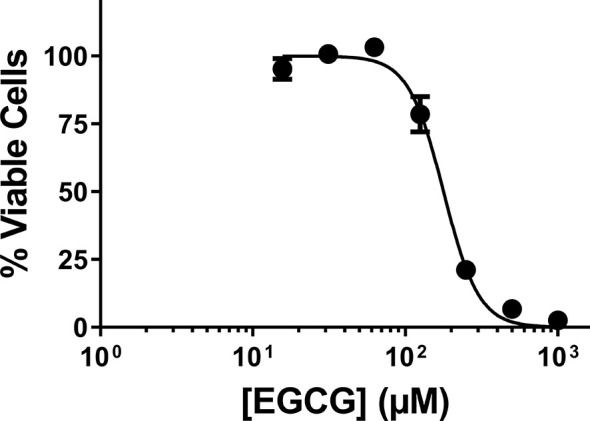
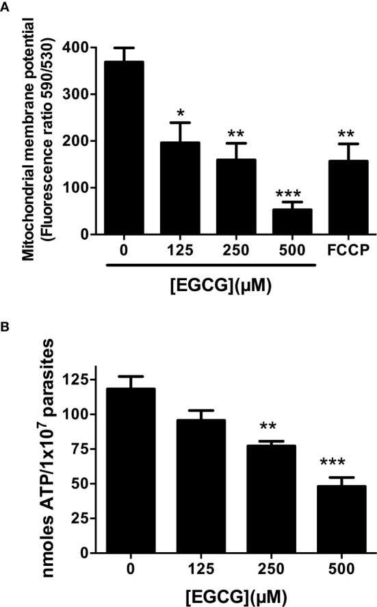
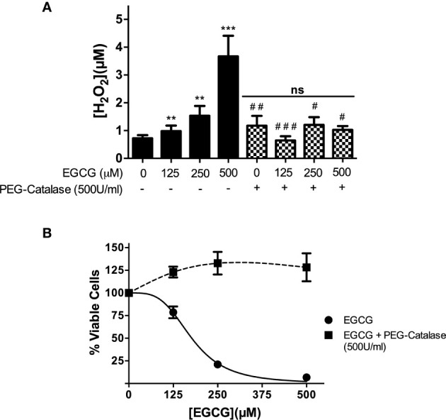
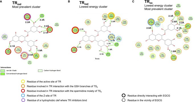
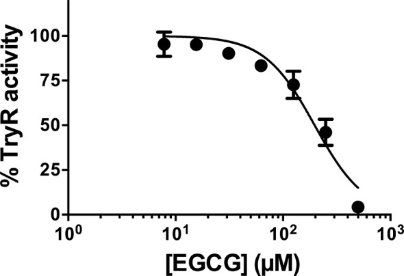
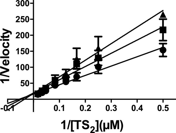
Leishmania infantum is a protozoan parasite that causes a vector borne infectious disease in humans known as visceral leishmaniasis (VL). This pathology, also caused by L. donovani, presently impacts the health of 500,000 people worldwide, and is treated with outdated anti-parasitic drugs that suffer from poor treatment regimens, severe side effects, high cost and/or emergence of resistant parasites. In previous works we have disclosed the anti-Leishmania activity of (-)-Epigallocatechin 3-O-gallate (EGCG), a flavonoid compound present in green tea leaves. To date, the mechanism of action of EGCG against Leishmania remains unknown. This work aims to shed new light into the leishmanicidal mode of action of EGCG. Towards this goal, we first confirmed that EGCG inhibits L. infantum promastigote proliferation in a concentration-dependent manner. Second, we established that the leishmanicidal effect of EGCG was associated with i) mitochondria depolarization and ii) decreased concentration of intracellular ATP, and iii) increased concentration of intracellular H2O2. Third, we found that the leishmanicidal effect and the elevated H2O2 levels induced by of EGCG can be abolished by PEG-catalase, strongly suggesting that this flavonoid kills L. infantum promastigotes by disturbing their intracellular redox balance. Finally, we gathered in silico and in vitro evidence that EGCG binds to trypanothione reductase (TR), a central enzyme of the redox homeostasis of Leishmania, acting as a competitive inhibitor of its trypanothione substrate.

Keywords: EGCG; Leishmania infantum; competitive inhibitor; mechanism of action; trypanothione reductase.


Conflict of interest statement

The authors declare that the research was conducted in the absence of any commercial or financial relationships that could be construed as a potential conflict of interest.

Figures

Figure 1
Figure 1
Effect of EGCG on Leishmania infantum. Promastigotes of L. infantum were cultivated in Schneider’s Drosophila medium at 26°C for 72 h in the absence or presence of EGCG (0.015 – 1 mM). Cell viability was measured using the alamar Blue assay. In the control (absence of EGCG), the same volume of PBS (solvent of EGCG) was added to the growth medium of controls. Values represent mean ± standard error of three different experiments.
Figure 2
Figure 2
EGCG induces mitochondrial depolarization in L. infantum with concomitant loss of intracellular ATP. Leishmania infantum promastigotes were cultivated in Schneider’s Drosophila medium at 26°C for 72 h in the absence or presence of 125 – 500 µM EGCG. Promastigotes were stained with the potentiometric probe JC-1 (10 µg/ml). The positive control was treated with FCCP (20 µM) for 20 minutes. In the control (absence of EGCG), the same volume of vehicle (PBS) was added to the growth medium. Concentration-dependent alterations in relative mitochondrial membrane potential (ΔΨm) values are expressed as the ratio of the fluorescence measurements at 590 nm (for J-aggregate) versus 530 nm (for J-monomer) (A). Promastigotes were incubated with EGCG (125 - 500 µM) for 72 h. Intracellular ATP concentrations were measured using a bioluminescence assay. The results are expressed as ATP concentration (B). Data represent means ± standard errors of three different experiments run in triplicate. * indicates a significant difference relative to the control group (p < 0.05); ** indicates a significant difference relative to the control group (p < 0.01); *** indicates a significant difference relative to the control group (p < 0.001).
Figure 3
Figure 3
EGCG induces a boost of H2O2 in L. infantum promastigotes that leads to parasite death. Promastigotes of L. infantum were cultivated in Schneider’s Drosophila medium at 26°C for 72 h with EGCG (125–500 µM) in the absence or in the presence of PEG-catalase (500 U/ml). H2O2 was measured with Amplex Red and expressed as the H2O2 concentration (A). Cellular viability was measured using the alamar Blue assay (B). Values represent mean ± standard error of three different experiments. ** indicates a significant difference relative to the control group (p < 0.01); *** indicates a significant difference relative to the control group (p < 0.001); # indicates a significant difference relative to the L. infantum incubated with 500 µM of EGCG (p < 0.05); ## indicates a significant difference relative to the L. infantum incubated with 500 µM of EGCG (p < 0.01); ### indicates a significant difference relative to the L. infantum incubated with 500 µM of EGCG (p < 0.001).
Figure 4
Figure 4
High confidence in silico models predict EGCG binding to the trypanothione binding site of trypanothione reductase (TR). Two dimensional representation of the interaction between EGCG (central carbon structure) and TR residues (green circles with aminoacid numbering; the letters A and B refer to different monomers) in (A) the most prevalent and also lowest energy EGCG/TRox cluster, (B) the lowest energy EGCG/TRred cluster, and (C) the most prevalent EGCG/TRred cluster.
Figure 5
Figure 5
L. infantum recombinant TR activity was inhibited by EGCG. L. infantum recombinant TR activity was assayed at 25°C and pH 7.5 in the presence of increasing concentrations of EGCG (7.8 – 500 µM), and the assays were carried out in triplicate. Control experiments were carried out in the absence of the inhibitor. Values represent mean ± standard error of three different experiments run in triplicate.
Figure 6
Figure 6
Kinetic analysis of TR activity in the presence of EGCG. The enzyme concentration was maintained as a constant, while the substrate [T(S)2] varied as described in the abscissa, and EGCG varied as follows: no addition (closed circles); 100 µM EGCG (closed squares); 200 µM EGCG (closed triangles). The values are presented as the mean ± standard error of three different experiments run in triplicate.

Similar articles

Cited by

References

    1. Amato V. S., Tuon F. F., Bacha H. A., Neto V. A., Nicodemo A. C. (2008). Mucosal leishmaniasis . Current scenario and prospects for treatment. Acta Trop. 105 (1), 1–9. 10.1016/j.actatropica.2007.08.003 - DOI - PubMed
    1. Baiocco P., Colotti G., Franceschini S., Ilari A. (2009. a). Molecular basis of antimony treatment in leishmaniasis. J. Med. Chem. 52 (8), 2603–2612. 10.1021/jm900185q - DOI - PubMed
    1. Baiocco P., Franceschini S., Ilari A., Colotti G. (2009. b). Trypanothione reductase from Leishmania infantum: cloning, expression, purification, crystallization and preliminary X-ray data analysis. Protein Pept. Lett. 16 (2), 196–200. 10.2174/092986609787316306 - DOI - PubMed
    1. Baiocco P., Poce G., Alfonso S., Cocozza M., Porretta G. C., Colotti G., et al. . (2013). Inhibition of Leishmania infantum trypanothione reductase by azole-based compounds: A comparative analysis with its physiological substrate by x-ray crystallography. ChemMedChem 8, 1175–1183. 10.1002/cmdc.201300176 - DOI - PubMed
    1. Barral A., Pedral-Sampaio D., Grimaldi Junior G., Momen H., McMahon-Pratt D., Ribeiro de Jesus A., et al. . (1991). Leishmaniasis in Bahia, Brazil: evidence that Leishmania amazonensis produces a wide spectrum of clinical disease. Am. J. Trop. Med. Hyg. 44 (5), 536–546. 10.4269/ajtmh.1991.44.536 - DOI - PubMed

Publication types

LinkOut - more resources