

Mitochondrial OMA1 and OPA1 as Gatekeepers of Organellar Structure/Function and Cellular Stress Response
- PMID: 33842456
- PMCID: PMC8027119
- DOI: 10.3389/fcell.2021.626117
Mitochondrial OMA1 and OPA1 as Gatekeepers of Organellar Structure/Function and Cellular Stress Response
Abstract
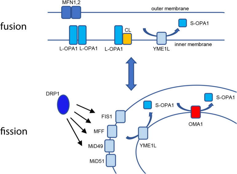
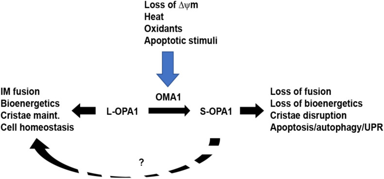
Mammalian mitochondria are emerging as a critical stress-responsive contributor to cellular life/death and developmental outcomes. Maintained as an organellar network distributed throughout the cell, mitochondria respond to cellular stimuli and stresses through highly sensitive structural dynamics, particularly in energetically demanding cell settings such as cardiac and muscle tissues. Fusion allows individual mitochondria to form an interconnected reticular network, while fission divides the network into a collection of vesicular organelles. Crucially, optic atrophy-1 (OPA1) directly links mitochondrial structure and bioenergetic function: when the transmembrane potential across the inner membrane (ΔΨm) is intact, long L-OPA1 isoforms carry out fusion of the mitochondrial inner membrane. When ΔΨm is lost, L-OPA1 is cleaved to short, fusion-inactive S-OPA1 isoforms by the stress-sensitive OMA1 metalloprotease, causing the mitochondrial network to collapse to a fragmented population of organelles. This proteolytic mechanism provides sensitive regulation of organellar structure/function but also engages directly with apoptotic factors as a major mechanism of mitochondrial participation in cellular stress response. Furthermore, emerging evidence suggests that this proteolytic mechanism may have critical importance for cell developmental programs, particularly in cardiac, neuronal, and stem cell settings. OMA1's role as a key mitochondrial stress-sensitive protease motivates exciting new questions regarding its mechanistic regulation and interactions, as well as its broader importance through involvement in apoptotic, stress response, and developmental pathways.
Keywords: OMA1; OPA1; apoptosis; development; mitochondria.
Copyright © 2021 Gilkerson, De La Torre and St. Vallier.
Conflict of interest statement
The authors declare that the research was conducted in the absence of any commercial or financial relationships that could be construed as a potential conflict of interest.
Figures
Similar articles
-
OMA1-Mediated Mitochondrial Dynamics Balance Organellar Homeostasis Upstream of Cellular Stress Responses.Int J Mol Sci. 2024 Apr 22;25(8):4566. doi: 10.3390/ijms25084566. Int J Mol Sci. 2024. PMID: 38674151 Free PMC article. Review.
-
Differentiation activates mitochondrial OPA1 processing in myoblast cell lines.Mitochondrion. 2024 Sep;78:101933. doi: 10.1016/j.mito.2024.101933. Epub 2024 Jul 8. Mitochondrion. 2024. PMID: 38986925 Free PMC article.
-
A Disturbance in the Force: Cellular Stress Sensing by the Mitochondrial Network.Antioxidants (Basel). 2018 Sep 22;7(10):126. doi: 10.3390/antiox7100126. Antioxidants (Basel). 2018. PMID: 30249006 Free PMC article. Review.
-
Mitochondrial OPA1 cleavage is reversibly activated by differentiation of H9c2 cardiomyoblasts.Mitochondrion. 2021 Mar;57:88-96. doi: 10.1016/j.mito.2020.12.007. Epub 2020 Dec 29. Mitochondrion. 2021. PMID: 33383158 Free PMC article.
-
Mitochondrial membrane potential and oxidative stress interact to regulate Oma1-dependent processing of Opa1 and mitochondrial dynamics.FASEB J. 2024 Sep 30;38(18):e70066. doi: 10.1096/fj.202400313R. FASEB J. 2024. PMID: 39312414
Cited by
-
Mitochondrial translation is the primary determinant of secondary mitochondrial complex I deficiencies.iScience. 2024 Jul 19;27(8):110560. doi: 10.1016/j.isci.2024.110560. eCollection 2024 Aug 16. iScience. 2024. PMID: 39184436 Free PMC article.
-
Honokiol alleviated neurodegeneration by reducing oxidative stress and improving mitochondrial function in mutant SOD1 cellular and mouse models of amyotrophic lateral sclerosis.Acta Pharm Sin B. 2023 Feb;13(2):577-597. doi: 10.1016/j.apsb.2022.07.019. Epub 2022 Aug 10. Acta Pharm Sin B. 2023. PMID: 36873166 Free PMC article.
-
Functional transcriptome analysis revealed major changes in pathways affecting systems biology of Tharparkar cattle under seasonal heat stress.3 Biotech. 2024 Jul;14(7):177. doi: 10.1007/s13205-024-04018-2. Epub 2024 Jun 6. 3 Biotech. 2024. PMID: 38855148 Free PMC article.
-
The Role of Mitochondria in Mediation of Skeletal Muscle Repair.Muscles. 2023 Mar 24;2(2):119-163. doi: 10.3390/muscles2020011. Muscles. 2023. PMID: 40757564 Free PMC article. Review.
-
Mechanistic study of optic atrophy 1 in ischemia-reperfusion disease.J Mol Med (Berl). 2023 Feb;101(1-2):1-8. doi: 10.1007/s00109-022-02271-7. Epub 2022 Nov 23. J Mol Med (Berl). 2023. PMID: 36418744 Review.
References
-
- Acin-Perez R., Lechuga-Vieco A. V., Mar Munoz M. Del, Nieto-Arellano R., Torroja C., Sanchez-Cabo F., et al. (2018). Ablation of the stress protease OMA1 protects against heart failure in mice. Sci. Transl. Med. 10:434. - PubMed
Publication types
Grants and funding
LinkOut - more resources
Full Text Sources
Other Literature Sources