Review
. 2021 Mar 26:9:628991.
doi: 10.3389/fcell.2021.628991. eCollection 2021.

From Flies to Men: ROS and the NADPH Oxidase in Phagocytes

Affiliations
Review

From Flies to Men: ROS and the NADPH Oxidase in Phagocytes

Zohreh Mansoori Moghadam et al. Front Cell Dev Biol. .

Abstract

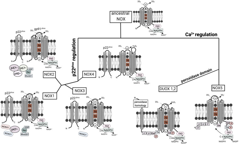
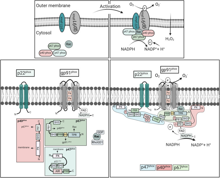
The cellular formation of reactive oxygen species (ROS) represents an evolutionary ancient antimicrobial defense system against microorganisms. The NADPH oxidases (NOX), which are predominantly localized to endosomes, and the electron transport chain in mitochondria are the major sources of ROS. Like any powerful immunological process, ROS formation has costs, in particular collateral tissue damage of the host. Moreover, microorganisms have developed defense mechanisms against ROS, an example for an arms race between species. Thus, although NOX orthologs have been identified in organisms as diverse as plants, fruit flies, rodents, and humans, ROS functions have developed and diversified to affect a multitude of cellular properties, i.e., far beyond direct antimicrobial activity. Here, we focus on the development of NOX in phagocytic cells, where the so-called respiratory burst in phagolysosomes contributes to the elimination of ingested microorganisms. Yet, NOX participates in cellular signaling in a cell-intrinsic and -extrinsic manner, e.g., via the release of ROS into the extracellular space. Accordingly, in humans, the inherited deficiency of NOX components is characterized by infections with bacteria and fungi and a seemingly independently dysregulated inflammatory response. Since ROS have both antimicrobial and immunomodulatory properties, their tight regulation in space and time is required for an efficient and well-balanced immune response, which allows for the reestablishment of tissue homeostasis. In addition, distinct NOX homologs expressed by non-phagocytic cells and mitochondrial ROS are interlinked with phagocytic NOX functions and thus affect the overall redox state of the tissue and the cellular activity in a complex fashion. Overall, the systematic and comparative analysis of cellular ROS functions in organisms of lower complexity provides clues for understanding the contribution of ROS and ROS deficiency to human health and disease.

Keywords: CGD; NADPH; inflammation; macrophages; mitochondrial ROS; myeloid cells; neutrophils; reactive oxygen species.


Conflict of interest statement

The authors declare that the research was conducted in the absence of any commercial or financial relationships that could be construed as a potential conflict of interest.

Figures

FIGURE 1
FIGURE 1
Structure and hypothetical evolution of NOX isoforms. The ancestral NOX enzyme consists of a conserved C-terminal core region including six transmembrane a-helices, two heme (Fe) groups, and a NADPH-binding cytoplasmic C-terminal domain. The conserved gp91phox subunit transfers electrons from NADPH to reduce oxygen (O2) to superoxide anion (O2). Later NOX isoforms acquired dependence on the activating subunit p22phox or calcium. NOX1–3 and NOX4 emerged from a common branch, as both of these subgroups depend on the presence of the p22phox subunit with two transmembrane a-helices. However, NOX4 solely relies on p22phox, while NOX1–3 require additional cytoplasmic subunits. Mammalian NOX2 additionally relies on the cytoplasmic regulators p47phox, p67phox, p40phox, and Rac. In contrast, NOX1 is regulated by NOXO1 and NOXA1. NOXO1 also contributes to NOX3 activation. NOX5 evolved from ancestral NOX through the acquisition of four EF hand motifs containing a Ca2+-binding domain, which enables activation by cytosolic calcium rather than other subunits. DUOX1/2 enzymes then emerged from NOX5 isoforms by the addition of a peroxidase domain in the N-terminal region enabling H2O2 production (Bedard et al., 2007; Kawahara et al., 2007).
FIGURE 2
FIGURE 2
Activation and assembly of mammalian NOX2. NOX2 consists of the cytosolic components p67phox, p47phox, p40phox, Rac2, and the integral membrane subunits gp91phox and p22phox. Upon cell stimulation, the cytosolic subunits translocate to the membranes to form an active complex with gp91phox and p22phox. Meanwhile, Rac exchanges GDP to GTP, and dissociates from Rho-GDI. In the resting state, the p47phox-SH3 tandem domain interacts with AIR keeping p47phox in an inactive conformation (Belambri et al., 2018). Cell stimulation induces phosphorylation of AIR, releasing the interactive domains, i.e., SH3, PX, and PRR, which mediate oxidase assembly. The PRR of p47phox binds to the SH3 region of p67phox, while p67phox links with p40phox through their PB1 domains. The p47phox-SH3 regions then bind to the p22phox-PRR domains promoting p67phox interaction with gp91phox and moving p40phox-PX domains in close proximity to the membrane. Activated NOX2 uses cytosolic NADPH to induce oxygen reduction and superoxide anion (O2⋅-) generation. Abbreviations: SH3, Src homology 3 (SH3);, PX, phox homology (PX);, AIR, auto-inhibitory region (AIR);, PRR, proline-rich region (PRR);, TPR, tetratricopeptide-rich regions; PB1, phox and Bem1 domain; and AD, activation domain.

Similar articles

Cited by

References

    1. Abuaita B. H., Schultz T. L., O’Riordan M. X. (2018). Mitochondria-Derived vesicles deliver antimicrobial reactive oxygen species to control phagosome-localized Staphylococcus aureus. Cell Host Microbe 24 625–636.e5. 10.1016/j.chom.2018.10.005 - DOI - PMC - PubMed
    1. Alimchandani M., Lai J.-P., Aung P. P., Khangura S., Kamal N., Gallin J. I., et al. (2013). Gastrointestinal histopathology in chronic granulomatous disease: a study of 87 patients. Am. J. Surg. Pathol. 37 1365–1372. 10.1097/PAS.0b013e318297427d - DOI - PMC - PubMed
    1. Antoniou E., Margonis G. A., Angelou A., Pikouli A., Argiri P., Karavokyros I., et al. (2016). The TNBS-induced colitis animal model: an overview. Ann. Med. Surg. 11 9–15. 10.1016/j.amsu.2016.07.019 - DOI - PMC - PubMed
    1. Arnadottir G. A., Norddahl G. L., Gudmundsdottir S., Agustsdottir A. B., Sigurdsson S., Jensson B. O., et al. (2018). A homozygous loss-of-function mutation leading to CYBC1 deficiency causes chronic granulomatous disease. Nat. Commun. 9:4447. 10.1038/s41467-018-06964-x - DOI - PMC - PubMed
    1. Arnold D. E., Heimall J. R. (2017). A review of chronic granulomatous disease. Adv. Ther. 34 2543–2557. 10.1007/s12325-017-0636-2 - DOI - PMC - PubMed

LinkOut - more resources