. 2021 Mar 25:9:660571.
doi: 10.3389/fcell.2021.660571. eCollection 2021.

Metallic Ions Encapsulated in Electrospun Nanofiber for Antibacterial and Angiogenesis Function to Promote Wound Repair

Affiliations

Metallic Ions Encapsulated in Electrospun Nanofiber for Antibacterial and Angiogenesis Function to Promote Wound Repair

Chenxi Zhu et al. Front Cell Dev Biol. .

Retraction in

Abstract

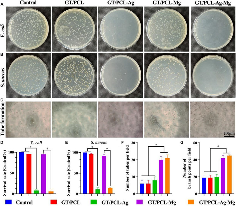
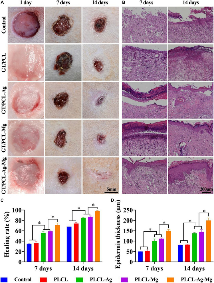
Electrospun nanofiber is an attractive biomaterial for skin tissue engineering because it mimics the natural fibrous extracellular matrix structure and creates a physical structure suitable for skin tissue regeneration. However, endowing the nanofibrous membranes with antibacterial and angiogenesis functions needs to be explored. In the current study, we aimed to fabricate gelatin/polycaprolactone (GT/PCL) (GT/PCL-Ag-Mg) nanofibers loaded with silver (Ag) and magnesium (Mg) ions for antibacterial activity and pro-angiogenesis function for wound repair. The fabricated GT/PCL membranes had a nanofibrous structure with random arrangement and achieved sustained release of Ag and Mg ions. In vitro results indicated that the GT/PCL-Ag-Mg membranes presented satisfactory cytocompatibility with cell survival and proliferation. In addition, the membranes with Ag demonstrated good antibacterial capacity to both gram-positive and gram-negative bacteria, and the Mg released from the membranes promoted the tube formation of vascular endothelial cells. Furthermore, in vivo results demonstrated that the GT/PCL-Ag-Mg membrane presented an accelerated wound healing process compared with GT/PCL membranes incorporated with either Ag or Mg ions and pure GT/PCL alone. Superior epidermis formation, vascularization, and collagen deposition were also observed in GT/PCL-Ag-Mg membrane compared with the other membranes. In conclusion, a multifunctional GT/PCL-Ag-Mg membrane was fabricated with anti-infection and pro-angiogenesis functions, serving as a potential metallic ion-based therapeutic platform for applications in wound repair.

Keywords: angiogenesis; antibacterial; electrospinning; magnesium; silver; wound healing.


Conflict of interest statement

The authors declare that the research was conducted in the absence of any commercial or financial relationships that could be construed as a potential conflict of interest.

Figures

SCHEME 1
SCHEME 1
The overall experimental design. A GT/PCL nanofiber membrane with Ag and Mg ions (GT/PCL-Ag-Mg) was fabricated, and its antibacterial and angiogenesis function were verified using in vitro and in vivo studies.
FIGURE 1
FIGURE 1
Characterizations of the nanofibrous membranes. (A) Gross and (B) SEM images of the fabricated GT/PCL, GT/PCL-Ag, GT/PCL-Mg, and GT/PCL-Ag-Mg membranes. (C) Mapping of carbon, Ag, and Mg elements in GT/PCL-Ag-Mg membrane. (D) FTIR analysis for Ag and Mg ions, as well as GT/PCL, GT/PCL-Ag, GT/PCL-Mg, and GT/PCL-Ag-Mg membranes. The releasing rate of (E) Ag and (F) Mg in the GT/PCL-Ag-Mg membrane.
FIGURE 2
FIGURE 2
Cytocompatibility of nanofibrous membranes. (A) Live and dead staining of fibroblasts cultured in vitro and (B) proliferation indicated by CCK-8 on Control, GT/PCL, GT/PCL-Ag, GT/PCL-Mg, and GT/PCL-Ag-Mg groups at 1 and 4 days. NS, no statistical difference.
FIGURE 3
FIGURE 3
In vitro antibacterial and pro-angiogenesis properties evaluations. Amount of (A) E. coli and (B) S. aureus on agar plates after incubation for 24 h on Control, GT/PCL, GT/PCL-Ag, GT/PCL-Mg, and GT/PCL-Ag-Mg groups. (C) Tube formation of HUVECs in vitro on Control, GT/PCL, GT/PCL-Ag, GT/PCL-Mg, and GT/PCL-Ag-Mg groups. Survival rate of (D) E. coli and (E) S. aureus on each group. Statistical analyses of tube (F) formation numbers and (G) branch points for each group. p < 0.05.
FIGURE 4
FIGURE 4
Gross and HE staining evaluations of wound healing. (A) Gross observations of wound healing at 1, 7, and 14 days on the Control, GT/PCL, GT/PCL-Ag, GT/PCL-Mg, and GT/PCL-Ag-Mg groups. (B) HE staining of repaired tissue at 7 and 14 days on the Control, GT/PCL, GT/PCL-Ag, GT/PCL-Mg, and GT/PCL-Ag-Mg groups. Quantitative analyses of (C) wound healing rate at 7 and 14 days and (D) epidermis thickness of repaired tissue on Control, GT/PCL, GT/PCL-Ag, GT/PCL-Mg, and GT/PCL-Ag-Mg groups. p < 0.05.
FIGURE 5
FIGURE 5
Evaluation of the collagen regeneration. (A) Masson’s trichrome and (B) Sirius red staining of repaired skin tissue at 7 and 14 days on Control, GT/PCL, GT/PCL-Ag, GT/PCL-Mg, and GT/PCL-Ag-Mg groups. Collagen fiber percentage of repaired tissue at 7 and 14 days on Control, GT/PCL, GT/PCL-Ag, GT/PCL-Mg, and GT/PCL-Ag-Mg groups (C). Scale bar: 100 μm. p < 0.05.
FIGURE 6
FIGURE 6
In vivo antibacterial and pro-angiogenesis properties evaluations. (A) FISH staining of bacteria at 3 days on Control, GT/PCL, GT/PCL-Ag, GT/PCL-Mg, and GT/PCL-Ag-Mg groups. (B) CD31 staining of repaired skin tissue at 7 and 14 days on Control, GT/PCL, GT/PCL-Ag, GT/PCL-Mg, and GT/PCL-Ag-Mg groups. Quantitative analyses of (C) bacterial counts and (D) neovascularization in repaired tissue at 7 and 14 days on Control, GT/PCL, GT/PCL-Ag, GT/PCL-Mg, and GT/PCL-Ag-Mg groups. p < 0.05.

Similar articles

Cited by

References

    1. Aavani F., Khorshidi S., Karkhaneh A. (2019). A concise review on drug-loaded electrospun nanofibres as promising wound dressings. J. Med. Eng. Technol. 43 38–47. 10.1080/03091902.2019.1606950 - DOI - PubMed
    1. Albright V., Xu M., Palanisamy A., Cheng J., Stack M., Zhang B., et al. (2018). Micelle-coated, hierarchically structured nanofibers with dual-release capability for accelerated wound healing and infection control. Adv. Healthc. Mater 7:e1800132. 10.1002/adhm.201800132 - DOI - PMC - PubMed
    1. Amberg R., Elad A., Rothamel D., Fienitz T., Szakacs G., Heilmann S., et al. (2018). Design of a migration assay for human gingival fibroblasts on biodegradable magnesium surfaces. Acta Biomater. 79 158–167. 10.1016/j.actbio.2018.08.034 - DOI - PubMed
    1. Atieh T., Audoly G., Hraiech S., Lepidi H., Roch A., Rolain J. M., et al. (2013). Evaluation of the diagnostic value of fluorescent in situ hybridization in a rat model of bacterial pneumonia. Diagn. Microbiol. Infect. Dis. 76 425–431. 10.1016/j.diagmicrobio.2013.04.028 - DOI - PubMed
    1. Bi H., Li H., Zhang C., Mao Y., Nie F., Xing Y., et al. (2019). Stromal vascular fraction promotes migration of fibroblasts and angiogenesis through regulation of extracellular matrix in the skin wound healing process. Stem Cell Res. Ther. 10:302. 10.1186/s13287-019-1415-6 - DOI - PMC - PubMed

Publication types

LinkOut - more resources