Review
. 2021 Mar 25:8:639587.
doi: 10.3389/fmolb.2021.639587. eCollection 2021.

Easy Synthesis of Complex Biomolecular Assemblies: Wheat Germ Cell-Free Protein Expression in Structural Biology

Affiliations
Review

Easy Synthesis of Complex Biomolecular Assemblies: Wheat Germ Cell-Free Protein Expression in Structural Biology

Marie-Laure Fogeron et al. Front Mol Biosci. .

Abstract

Cell-free protein synthesis (CFPS) systems are gaining more importance as universal tools for basic research, applied sciences, and product development with new technologies emerging for their application. Huge progress was made in the field of synthetic biology using CFPS to develop new proteins for technical applications and therapy. Out of the available CFPS systems, wheat germ cell-free protein synthesis (WG-CFPS) merges the highest yields with the use of a eukaryotic ribosome, making it an excellent approach for the synthesis of complex eukaryotic proteins including, for example, protein complexes and membrane proteins. Separating the translation reaction from other cellular processes, CFPS offers a flexible means to adapt translation reactions to protein needs. There is a large demand for such potent, easy-to-use, rapid protein expression systems, which are optimally serving protein requirements to drive biochemical and structural biology research. We summarize here a general workflow for a wheat germ system providing examples from the literature, as well as applications used for our own studies in structural biology. With this review, we want to highlight the tremendous potential of the rapidly evolving and highly versatile CFPS systems, making them more widely used as common tools to recombinantly prepare particularly challenging recombinant eukaryotic proteins.

Keywords: NMR; cell-free protein expression; labeling; structural biology; wheat germ.


Conflict of interest statement

MH declares competing financial interests as an employee of CellFree Sciences Co., Ltd., Japan. The other authors do not have any competing interests.

Figures

FIGURE 1
FIGURE 1
Components of WG-CFPS described by Takai et al. (2010). (A) Wheat germ extract (WGE) can be prepared from nontreated durum wheat. Alternatively, commercially available WGE can be used. (B) The WG-CFPS uses expression templates having a SP6 promoter to drive RNA synthesis and an E01 translational enhancer to induce cap-independent translation. The system can use circular and linear DNA templates to hold a cDNA encoding a protein. Alternatively, T7 RNA polymerase can be used as well under the same reaction conditions. The RNA is used as a template for protein synthesis. (C) The key components for protein synthesis are provided with the WGE. This includes the necessary ribosomes and tRNAs, but there are also other cell components in those extracts that may assist for example protein folding or possibly protein modification. Other key components are provided by the “Buffered Substrate Solution” which includes the amino acids, a DTT-based redox system, and a creatine kinase driving energy supply. Protein synthesis reactions can be modified as further explained in the text. (D) Protein synthesis can be confirmed by several different methods with the most commonly ones given in the figure.
FIGURE 2
FIGURE 2
Workflow to establish protein synthesis in a WGS, with key points given at each step.
FIGURE 3
FIGURE 3
Template design for expression of the protein of interest. (A) Design of the gene sequence. (B) Both circular and linear DNA templates can be used for transcription.
FIGURE 4
FIGURE 4
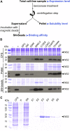
Protein analysis. (A) Typical flowchart for protein analysis after small-scale expression test. Parameters to be considered are highlighted in blue. (B) Small-scale expression test of the nonstructural protein 2 (NS2) from hepatitis C virus (HCV). This membrane protein was produced in the absence or presence of various detergents at a 0.1% concentration (w/v). Samples were analyzed by SDS-PAGE followed by Coomassie blue staining (upper panels) and Western blotting with an antibody against the Strep-tag II fused at the C-terminus of NS2 (lower panels). CFS, total cell-free sample; pellet, pellet obtained after centrifugation of CFS; SN-beads, supernatant obtained after centrifugation of CFS and incubated with Strep-Tactin magnetic beads to capture Strep-tag II-tagged NS2 protein; −, negative control (no NS2); +, positive control (NS2 expressed in the absence of detergent). The black arrowheads indicate NS2, adapted from Fogeron et al. (2015a). (C) SDS-PAGE analysis followed by Coomassie blue staining of the different steps from the affinity purification of the NS2 membrane protein produced directly in a solubilized form in the presence of MNG-3, adapted from Fogeron et al. (2015b).
FIGURE 5
FIGURE 5
Different reaction formats for protein expression using the WGS. (A) Schematic representation and (B) picture of a 500 μL dialysis cassette for medium scale CECF production. (C) Schematic representation of the bilayer method which is performed either in a 96-well plate for small-scale expression test or in a 6-well plate for larger-scale production (D), adapted from Fogeron et al. (2015a), Fogeron et al. (2015b); Fogeron et al. (2017b). (E) Schematic representation of the dialysis method and (F) picture of a CECF mini-reactor manufactured at ETH Zurich by Andreas Hunkeler in Beat H. Meier’s laboratory, according to Schneider et al. (2009). In this reaction format, a 24-well plate is used. For all panels, the translation mix is represented in yellow while the feeding buffer is represented in blue.
FIGURE 6
FIGURE 6
Structural characterization of proteins produced from cell-free protein expression.
FIGURE 7
FIGURE 7
Examples of structural studies on proteins expressed in WGS using NMR, X-ray crystallography, and cryo-EM. (A) Solid-state NMR spectrum of the HCV membrane protein NS4B reconstituted into DMPC lipids (Jirasko et al., 2020). (B) Dimer orientation in lipids of the HCV helix anchor and domain 1 (AHD1) of the NS5A protein as determined by solid-state NMR (Jirasko et al., 2020). (C) Solid-state NMR spectra of the hepatitis B virus capsid (Wang et al., 2019) and of (D) the subviral particles made of duck HBV small envelope protein (DHBs S) (David et al., 2018). The three spectra have been recorded at 110 kHz MAS on an 850 MHz spectrometer. Both HBV capsids and subviral particles were autoassembled during cell-free synthesis; their negative-staining electron microscopy images are shown inside the corresponding spectrum. (E) 20 conformers obtained by solution NMR of At3g01050.1 protein (Vinarov et al., 2004) (PDB 1se9, figure prepared using PyMoL (https://pymol.org/2/). (F) Structure of restriction endonuclease PabI obtained by X-ray crystallography (Miyazono et al., 2007; Watanabe et al., 2010) (PDB 2dvy). (G) 3D cryo-EM reconstruction of PDX1.2 complex at 15 Å resolution (Novikova et al., 2018). Figures were adapted with permission from Jirasko et al. (2020) for panel A, from Wang et al. (2019) for panel C, and from David et al. (2018) for panel D and reprinted with permission from Jirasko et al. (2020) for panel D, from Miyazono et al. (2007), Watanabe et al. (2010) for panel F, and from Novikova et al. (2018) for panel G.

References

    1. Abe M., Hori H., Nakanishi T., Arisaka F., Ogasawara T., Sawasaki T., et al. (2004). Application of cell-free translation systems to studies of cofactor binding proteins. Nucleic Acids Symp. Ser. 13, 143–144. 10.1093/nass/48.1.143 - DOI - PubMed
    1. Aceti D. J., Bingman C. A., Wrobel R. L., Frederick R. O., Makino S., Nichols K. W., et al. (2015). Expression platforms for producing eukaryotic proteins: a comparison of E. coli cell-based and wheat germ cell-free synthesis, affinity and solubility tags, and cloning strategies. J. Struct. Funct. Genomics 16, 67–80. 10.1007/s10969-015-9198-1 - DOI - PMC - PubMed
    1. Agarwal V., Penzel S., Szekely K., Cadalbert R., Testori E., Oss A., et al. (2014). De novo 3D structure determination from sub-milligram protein samples by solid-state 100 kHz MAS NMR spectroscopy. Angew. Chem. Int. Ed. Engl. 53, 12253–12256. 10.1002/anie.201405730 - DOI - PubMed
    1. Akbarzadeh A., Rezaei-Sadabady R., Davaran S., Joo S. W., Zarghami N., Hanifehpour Y., et al. (2013). Liposome: classification, preparation, and applications. Nanoscale Res. Lett. 8, 102. 10.1186/1556-276X-8-102 - DOI - PMC - PubMed
    1. Althoff T., Davies K. M., Schulze S., Joos F., Kühlbrandt W. (2012). GRecon: a method for the lipid reconstitution of membrane proteins. Angew. Chem. Int. Ed. Engl. 51, 8343–8347. 10.1002/anie.201202094 - DOI - PMC - PubMed

LinkOut - more resources