

Preparation of a Composite Scaffold from Polycaprolactone and Hydroxyapatite Particles by Means of Alternating Current Electrospinning
- PMID: 33842792
- PMCID: PMC8028135
- DOI: 10.1021/acsomega.1c00644
Preparation of a Composite Scaffold from Polycaprolactone and Hydroxyapatite Particles by Means of Alternating Current Electrospinning
Abstract
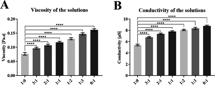
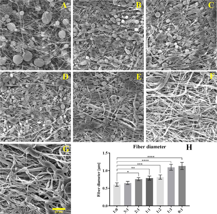
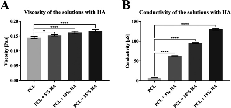
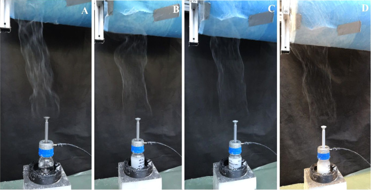
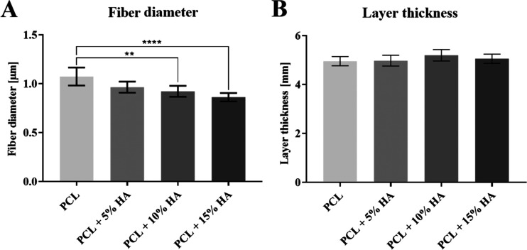
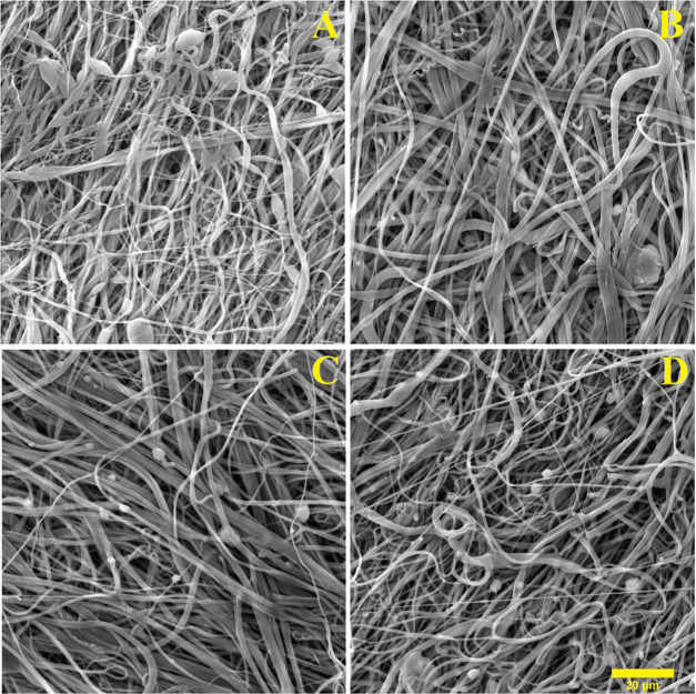
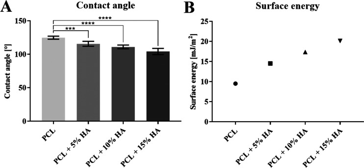
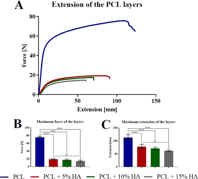
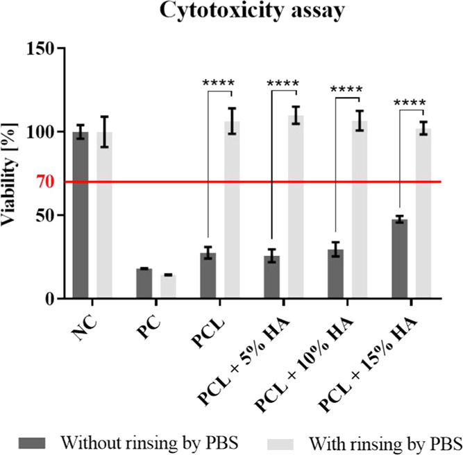
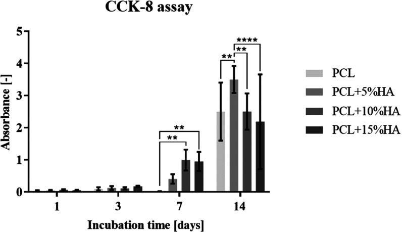
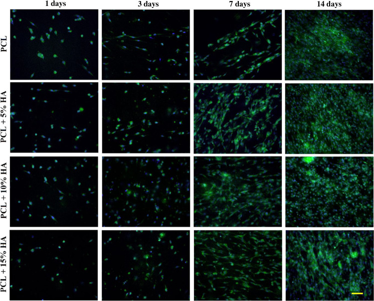
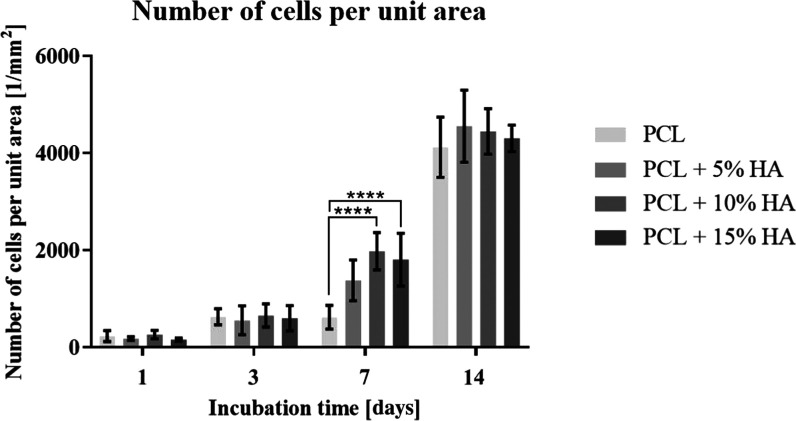
This research involved the production of polycaprolactone fiber layers via the alternating current electrospinning method. To construct the micro/nanofiber scaffold, mixtures of two molecular weight solutions, M n 45 000 and M n 80 000, were spun in differing proportions in a solvent system containing acetic acid, formic acid, and acetone in a ratio of 1:1:1. The composite fiber materials with hydroxyapatite particles were prepared from a solution that combined the different molecular weight solutions at a ratio of 1:3. The study resulted in the preparation of fiber layers containing 0, 5, 10, and 15% (wt) hydroxyapatite particles from the dry mass of the polycaprolactone. The strength, wettability, and surface energy of the composite materials were examined, and the results demonstrated that hydroxyapatite affects the fiber diameters, strength, and surface energy and, thus, the wettability of the fiber layers. The fibrous layers produced were further tested for cytotoxicity and cell viability and proliferation. The results obtained thus strongly indicate that the resulting bulky micro/nanofiber layers are suitable for further testing with a view to their eventual application in the field of bone tissue engineering.
© 2021 The Authors. Published by American Chemical Society.
Conflict of interest statement
The authors declare no competing financial interest.
Figures
References
-
- Nair N. R.; Sekhar V. C.; Nampoothiri K. M.; Pandey A.. 32—Biodegradation of Biopolymers. In Current Developments in Biotechnology and Bioengineering; Pandey A.; Negi S.; Soccol C. R., Eds.; Elsevier, 2017; pp 739–755.
-
- McKeen L.12—Renewable Resource and Biodegradable Polymers. In The Effect of Sterilization on Plastics and Elastomers; 3rd ed.; McKeen L., Ed.; William Andrew Publishing: Boston, 2012; pp 305–317.
-
- Erben J.; Jencova V.; Chvojka J.; Blazkova L.; Strnadova K.; Modrak M.; Kostakova E. K. The Combination of Meltblown Technology and Electrospinning—The Influence of the Ratio of Micro and Nanofibers on Cell Viability. Mater. Lett. 2016, 173, 153–157. 10.1016/j.matlet.2016.02.147. - DOI
-
- Klicova M.; Klapstova A.; Chvojka J.; Koprivova B.; Jencova V.; Horakova J. Novel Double-Layered Planar Scaffold Combining Electrospun PCL Fibers and PVA Hydrogels with High Shape Integrity and Water Stability. Mater. Lett. 2020, 263, 12728110.1016/j.matlet.2019.127281. - DOI
LinkOut - more resources
Full Text Sources
Other Literature Sources
