Review
. 2021 Jan 18;4(1):163-194.
doi: 10.1021/acsabm.0c01139. Epub 2020 Dec 23.

Biodegradable Materials for Sustainable Health Monitoring Devices

Affiliations
Review

Biodegradable Materials for Sustainable Health Monitoring Devices

Ensieh S Hosseini et al. ACS Appl Bio Mater. .

Abstract

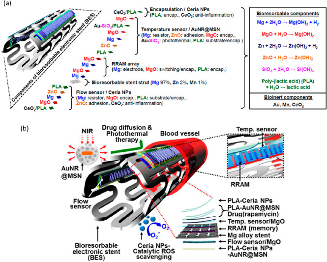
The recent advent of biodegradable materials has offered huge opportunity to transform healthcare technologies by enabling sensors that degrade naturally after use. The implantable electronic systems made from such materials eliminate the need for extraction or reoperation, minimize chronic inflammatory responses, and hence offer attractive propositions for future biomedical technology. The eco-friendly sensor systems developed from degradable materials could also help mitigate some of the major environmental issues by reducing the volume of electronic or medical waste produced and, in turn, the carbon footprint. With this background, herein we present a comprehensive overview of the structural and functional biodegradable materials that have been used for various biodegradable or bioresorbable electronic devices. The discussion focuses on the dissolution rates and degradation mechanisms of materials such as natural and synthetic polymers, organic or inorganic semiconductors, and hydrolyzable metals. The recent trend and examples of biodegradable or bioresorbable materials-based sensors for body monitoring, diagnostic, and medical therapeutic applications are also presented. Lastly, key technological challenges are discussed for clinical application of biodegradable sensors, particularly for implantable devices with wireless data and power transfer. Promising perspectives for the advancement of future generation of biodegradable sensor systems are also presented.


Conflict of interest statement

The authors declare no competing financial interest.

Figures

Figure 1
Figure 1
Chemical structures of natural and synthetic biodegradable polymeric materials. Chemical structures of moieties susceptible to hydrolysis and oxidation. Mechanism of acid-catalyzed ester hydrolysis. Mechanism of PVA enzymatic degradation. Reproduced with permission from refs ( and 58). Copyright 2008 Woodhead Publishing and ref (59). Copyright 2014 WILEY-VCH Verlag GmbH and Co. KGaA, Weinheim.
Figure 2
Figure 2
(a) Ultrathin devices on a flexible silk substrate, in flat (left) and bent (center and right) configurations, and (b) images of the water dissolution of silicon electronics on silk, at various time stages. Reproduced with permission from ref (19). Copyright 2009 American Institute of Physics (AIP). (c) Photographs of plasticized silk electrodes conformably attached to a finger and laminated on a human forearm. Reproduced with permission from ref (97). Copyright 2018 WILEY-VCH Verlag GmbH and Co. KGaA, Weinheim. (d) Photographs showing a silk-graphene E-tattoo attached to the forearm and the tattoo on skin in a stretched (upper), compressed (middle), and twisted (lower) state. Reproduced with permission from ref (98). Copyright 2019 WILEY-VCH Verlag GmbH and Co. KGaA, Weinheim.
Figure 3
Figure 3
(a) Schematic diagram demonstrating the fabrication of a noninvasive periodontal diagnostic sensor, a breath analyzer, and a gesture sensor using NiSe2 modified cellulose paper. Reproduced with permission from ref (103). Copyright 2019 American Chemical Society. (b) Schematic diagram demonstrating the fabrication of a real-time paper-based H2O2 sensing chip and image of a respiratory mask that includes customized sidewalls and an extension of a commercial filter with the paper-based sensor. Reproduced with permission from ref (104). Copyright 2019 American Chemical Society. (c) Integration of a 3D stacked paper-based autonomous healthcare monitoring system. Reproduced with permission from ref (105). Copyright 2017 WILEY-VCH Verlag GmbH and Co. KGaA, Weinheim. (d) Paper-based sweat sensor for human perspiration monitoring. Reproduced with permission from ref (106). Copyright 2019 WILEY-VCH Verlag GmbH and Co. KGaA, Weinheim.
Figure 4
Figure 4
(a) Photographs of a flexible and transparent starch–chitosan substrate (SC) and dissolution of an SC based transparent electrode. Reproduced with permission from ref (113). Copyright 2018 American Chemical Society. (b) Photographs of a chitosan–PVP substrate and its biodegradability in soil. Reproduced with permission from ref (115). Copyright 2019 American Chemical Society.
Figure 5
Figure 5
(a) Schematic diagram demonstrating the fabrication of an OFET device using a three-arm stereocomplex PLA substrate and images of the fully constructed transparent and flexible OFETs. Reproduced with permission from ref (132). Copyright 2015 WILEY-VCH Verlag GmbH and Co. KGaA, Weinheim. (b) Photographs of a flexible wearable transient MXene/tissue paper sensor sandwiched between a PLA thin sheet and an interdigitated electrode-coated PLA thin sheet, and its dissolution over 2 weeks in a 0.5 M NaOH solution. Reproduced with permission from ref (133). Copyright 2019 American Chemical Society. (c) Sequential dissolution images of a fully transient PVA-based electrochemical strip in DI water. Reproduced with permission from ref (135). Copyright 2020 The Royal Society of Chemistry. (d) Photographs of a PVA–liquid metal hydrogel for wearable transient epidermal sensors placed in a HCl solution. Reproduced with permission from ref (136). Copyright 2019 American Chemical Society. (e) Images showing the degradation of a patterned electrospun nanofibrous PGS–PCL substrate inside a solution of 0.5 M NaOH. Reproduced with permission from ref (127). Copyright 2014 WILEY-VCH Verlag GmbH and Co. KGaA, Weinheim.
Figure 6
Figure 6
(a) Schematic diagram representing the wafer-scale fabrication of fully formed transient n-channel MOSFETs based on SiO2 gate and interlayer dielectrics, and subsequent transfer of the device to silk films. (b) Optical images of the dissolution and disintegration of an array of MOSFETs on silk. Reproduced with permission from ref (142). Copyright 2013 WILEY-VCH Verlag GmbH and Co. KGaA, Weinheim. (c) Images of the degradation of a highly sensitive biodegradable pressure sensor based on nanofibrous PLGA–PCL dielectric in PBS. Reproduced with permission from ref (147). Copyright 2019 Elsevier. (d) Rollable pentacene OTFT with silk fibroin as the gate dielectric. Reproduced with permission from ref (165). Copyright 2011 WILEY-VCH Verlag GmbH and Co. KGaA, Weinheim. (e) Schematic diagram representing the structure of an OFET fabricated using egg white as a dielectric and a schematic diagram demonstrating the denaturation and cross-linking of albumen protein under thermal treatment. Reproduced with permission from ref (178). Copyright 2011 WILEY-VCH Verlag GmbH and Co. KGaA, Weinheim.
Figure 7
Figure 7
(a) Image of the fabrication and degradation of capacitive electrophysiology (EP) sensor based on biodegradable elastomers and Si nanomembranes/nanoribbons. Reproduced with permission from ref (180). Copyright 2015 American Chemical Society. (b) Illustration and chemical structure of nanoconfined acid-labile semiconductor fibers embedded within a biodegradable elastomer as well as the UV–vis absorption spectra of a solution of p(DPP-PPD) chlorobenzene with the addition of 1% 1 M TFA, and the normalized peak maxima extracted from UV–vis absorption spectra of a thin film of neat and nanoconfined p(DPP-PPD) in 1 M TFA water. Reproduced with permission from ref. Copyright 2019 American Chemical Society.
Figure 8
Figure 8
Time-lapse images of the dissolution of Mg electrodes of chitosan-based resistive switching memory devices on a plastic substrate in water at room temperature. Reproduced with permission from ref (221). Copyright 2015 WILEY-VCH Verlag GmbH and Co. KGaA, Weinheim.
Figure 9
Figure 9
(a) Epidermal electronic systems mounted on a forearm and encapsulated with a layer of spray-on-bandage (left), under compression and extension of the skin (center), and after wearing for 1 week (right). Reproduced with permission from ref (225). Copyright 2013 WILEY-VCH Verlag GmbH and Co. KGaA, Weinheim. (b) Monocrystalline silicon nanomembranes as encapsulation layers for water-soluble electronics. Reproduced with permission from ref (64). Copyright 2017 American Chemical Society.
Figure 10
Figure 10
(a) Schematic cross-section of a passive LC resonant sensor and (b) capacitor design. Reproduced with permission from ref (231). Copyright 2014 IEEE. (c, d) Schematic and optical image of the biodegradable piezoelectric PLLA sensor. (e) Sensor degradation at different days in the buffered solution at a temperature of 74 °C. Reproduced with permission from ref (232). Copyright 2018 Proceedings of the National Academy of Sciences of the United States of America.
Figure 11
Figure 11
(a) Schematic illustration of the structure of the biodegradable and elastomer PGS-based sensor. (b) SEM image of the PGS film contains microstructured 2D arrays of square-pyramid shapes. (c) Photographs of a device before in vitro degradation and after 7 weeks of incubation. Reproduced with permission from ref (234). Copyright 2015 WILEY-VCH Verlag GmbH and Co. KGaA, Weinheim. (d–f) Sketch of the structure and functional principle of the wirelessly readable capacitive pressure sensor. Reproduced with permission from ref (230). Copyright 2019 The Author(s), under exclusive license to Springer Nature Limited. (g) Implantable strain and pressure sensor that can be attached to a tendon for real-time healing assessment. (h) Materials and overall assembly of the fully biodegradable sensor. Reproduced with permission from ref (137). Copyright 2018 Macmillan Publishers Limited, part of Springer Nature.
Figure 12
Figure 12
(a) Schematic and optical image of the biodegradable piezoresistive PLLA sensor consists of Si-NM strain gauge as a sensing element. (b) Schematic illustration of the working principle and (c) pressure response performance of the biodegradable sensors (blue line) and its comparison with commercial sensor (orange line). Reproduced with permission from ref (235). Copyright 2020 WILEY-VCH Verlag GmbH and Co. KGaA, Weinheim. (d) Schematic illustration of the piezoresistive silicon-nanomembrane (Si-NM) pressure sensor. The inset shows the magnified illustration of the Si-NM strain gauge. (e) Photographs of the device dissolution upon insertion into an aqueous buffer solution (pH 12) at room temperature. Reproduced with permission from ref (1). Copyright 2016 Nature Publishing Group.
Figure 13
Figure 13
Optical images of the (a) glycine/chitosan (Gly/CS) film and (b, c) deposited Au electrodes on film and fabricated flexible sensor. (d) Dissolution of biodegradable piezoelectric Gly/CS-based pressure sensor with Mg electrodes in PBS solution over time. Reproduced with permission from ref (7). Copyright 2020 American Chemical Society.
Figure 14
Figure 14
(a) Schematic structure and optical image of resistive temperature sensor consist of metal and semiconductor serpentine structure as sensing element, Peano-like interconnects, and Ecoflex encapsulation. Biodegradability of the sensor in a water–NaCl solution (b) after 36 d and (c) 67 d dissolution in water. (d) Changes in resistance of the sensor after 24 h immersing in the solution. Reproduced with permission from ref (243). Copyright 2017 WILEY-VCH Verlag GmbH and Co. KGaA, Weinheim.
Figure 15
Figure 15
Composite temperature sensors using different sensing materials. (a) Image of a fibrous temperature sensor. (b) Three different conducting mechanisms for composite CNT temperature sensors: (i–iii) Three different electron/ions transport modes in CNT and [EMIM]Tf2N composite sensing materials: (i) transport of charges via the body–body contacts among CNTs; (ii) transport via the end to end contacts of CNTs; (iii) transport via ionic liquids. (c, d) Scanning electron microscopic (SEM) images of the cross-section of a fibrous temperature sensor in different scales: Ecoflex constitutes the outside sealing layer, silk fibers encapsulated with CNTs and [EMIM]Tf2N turn out to be the middle layer, and polyester fibers were chosen as the supporting cores. (e) SEM displays the surface morphology of composite material of CNTs and [EMIM]Tf2N between the middle silk coiling fibers. Reproduced with permission from ref (244). Copyright 2019 WILEY-VCH Verlag GmbH and Co. KGaA, Weinheim.
Figure 16
Figure 16
(a) Photo of an LIG electronic circuit on a thin leaf for flexible and wearable devices. The inset shows an enlarged optical image of the temperature sensor (scale bar: 1 mm). (b) Photo of LIG electronics on a leaf for green electronics. (c) Photo of the LIG temperature sensor on the leaf and an experimental setup for measuring performance. The inset shows the flexibility of the leaf that was still retained after FsLDW. (d) Resistance variation as a function of temperature, indicating a negative temperature coefficient behavior. The inset shows the dependency of ln(R) on 1/T. Reproduced with permission from ref (245). Copyright 2019 WILEY-VCH Verlag GmbH and Co. KGaA, Weinheim.
Figure 17
Figure 17
(a) Components of a bioresorbable electronic stent (BES) and bioinert materials and (b) schematic diagram of the BES that includes Mg alloy stent integrated with ceria NPs (catalytic ROS scavenging), AuNR@MSN (photothermal therapy), drugs (e.g., Rapamycin; well-known drug for the treatment of restenosis), flow/temperature sensors (physiological signal sensing), and RRAM array (data storage). Reproduced with permission from ref (246). Copyright 2015 American Chemical Society.

References

    1. Kang S.-K.; Murphy R. K.; Hwang S.-W.; Lee S. M.; Harburg D. V.; Krueger N. A.; Shin J.; Gamble P.; Cheng H.; Yu S.; et al. Bioresorbable silicon electronic sensors for the brain. Nature 2016, 530 (7588), 71–76. 10.1038/nature16492. - DOI - PubMed
    1. Tao H.; Hwang S.-W.; Marelli B.; An B.; Moreau J. E.; Yang M.; Brenckle M. A.; Kim S.; Kaplan D. L.; Rogers J. A.; et al. Silk-based resorbable electronic devices for remotely controlled therapy and in vivo infection abatement. Proc. Natl. Acad. Sci. U. S. A. 2014, 111 (49), 17385–17389. 10.1073/pnas.1407743111. - DOI - PMC - PubMed
    1. Dahiya R. E-Skin: From humanoids to humans. Proc. IEEE 2019, 107 (2), 247–252. 10.1109/JPROC.2018.2890729. - DOI
    1. Manjakkal L.; Dang W.; Yogeswaran N.; Dahiya R. Textile-based potentiometric electrochemical pH sensor for wearable applications. Biosensors 2019, 9 (1), 14. 10.3390/bios9010014. - DOI - PMC - PubMed
    1. Kim D.-H.; Lu N.; Ma R.; Kim Y.-S.; Kim R.-H.; Wang S.; Wu J.; Won S. M.; Tao H.; Islam A.; Yu K. J.; Kim T.-i.; Chowdhury R.; Ying M.; Xu L.; Li M.; Chung H.-J.; Keum H.; McCormick M.; Liu P.; Zhang Y.-W.; Omenetto F. G.; Huang Y.; Coleman T.; Rogers J. A. Epidermal Electronics. Science 2011, 333 (6044), 838. 10.1126/science.1206157. - DOI - PubMed

Publication types