. 2021 Mar 25:4:630955.
doi: 10.3389/frai.2021.630955. eCollection 2021.

Conformational Changes of the Receptor Binding Domain of SARS-CoV-2 Spike Protein and Prediction of a B-Cell Antigenic Epitope Using Structural Data

Affiliations

Conformational Changes of the Receptor Binding Domain of SARS-CoV-2 Spike Protein and Prediction of a B-Cell Antigenic Epitope Using Structural Data

Sangeeta Khare et al. Front Artif Intell. .

Abstract

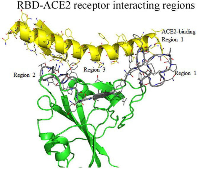
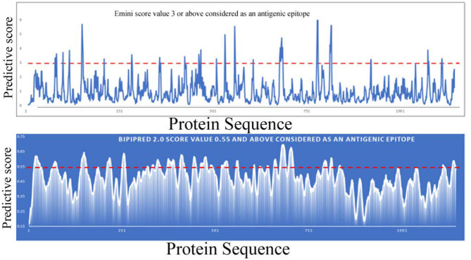
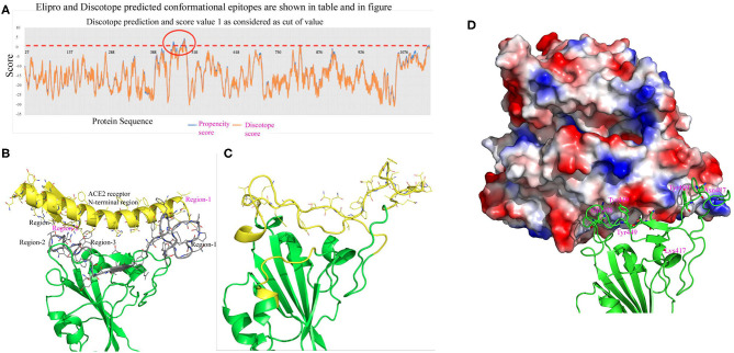
COVID-19, the illness caused by the SARS-CoV-2 virus, is now a worldwide pandemic with mortality in hundreds of thousands as infections continue to increase. Containing the spread of this viral infection and decreasing the mortality rate is a major challenge. Identifying appropriate antigenic epitopes from the viral proteins is a very important task for vaccine production and the development of diagnostic kits and antibody therapy. A novel antigenic epitope would be specific to the SARS-CoV-2 virus and can distinguish infections caused by common cold viruses. In this study two approaches are employed to identify both continuous and conformational B-cell antigenic epitopes. To achieve this goal, we modeled a complete structure of the receptor binding domain (RBD) of the spike protein using recently deposited coordinates (6vxx, 6vsb, and 6w41) in the protein data bank. In addition, we also modeled the RBD-ACE2 receptor complex for SARS-CoV-2 using the SARS-CoV RBD-ACE2 complex (3D0J) as a reference model. Finally, structure based predicted antigenic epitopes were compared to the ACE2 binding region of RBD of SARS-CoV-2. The identified conformational epitopes show overlaps with the ACE2-receptor binding region of the RBD of SARS-CoV-2. Strategies defined in the current study identified novel antigenic epitope that is specific to the SARS-CoV-2 virus. Integrating such approach in the diagnosis can distinguish infections caused by common cold viruses from SARS-CoV-2 virus.

Keywords: SARS–CoV−2; conformational epitope; receptor binding domian; spike protein; viral infection.


Conflict of interest statement

The authors declare that the research was conducted in the absence of any commercial or financial relationships that could be construed as a potential conflict of interest.

Figures

Figure 1
Figure 1
(A) C-α atom superposition of 6vsb with 6vxx coordinates. This alignment shows C-α atoms from residues 27–318 and 563–1146 aligns on top of each other with root mean square deviation (rmsd) 0.86 Å. (B) Magnified cartoon diagram that shows only RBD of both structures with superposition and RMSD between these two structures is ~38 Å. The globular head shows the missing loops region in both structures and part of the region is shown in yellow color. Color code: Green color-closed form and red color-open form. (C) Structural superposition of the RBD of 6w41 coordinates on top of RBD of 6vxx and 6vsb. (D) Cartoon diagram shows the superposition of the RBD of 6w41 coordinates on top of both structural forms. Color code: Green color-closed form, red- (complete structure of RBD from pdb 6w41), and yellow (RBD lack loops) the open form. The distance between these two structures are also shown in dotted lines.
Figure 2
Figure 2
(A) Cartoon diagram shows that residues 324–328 form a loop instead of β-strand in open conformation. This cartoon representation shows the β-strands of RBD domain movements during the conformational change. The figure also shows that during open conformation the entire domain moves 10 Å upwards (shown by blue arrow) and β-strands rotational angles changes from 75 to 110° (see Table 1 for detail information). Color code: Green-closed form and Red-open form. (B,C) Electropotential surface of the RBD for closed and open conformations, respectively. It also shows structural rearrangement leads to change in the electropotential surface, which are highlighted by numbers (1 to 4). The rearrangements occur at clockwise direction during structural change. Right side top figure is for closed form and bottom is for open form.
Figure 3
Figure 3
Interaction between regions of RBD of SARS-CoV-2 and the ACE2 receptor molecule. The RBD globular head forms a bowl-shaped structure and both ends are covered with elevated loops. Residues from these loops are positioned to interact with the ACE2 receptors and labeled as ACE2 receptor binding region 1 to 3 and highlighted with gray color. The interacting residues of ACE2 receptor also labeled as region 1 to region 3.
Figure 4
Figure 4
Linear B-cell epitope prediction by various bioinformatics tools [Emini (upper panel), BepiPred-2.0 (lower panel)].
Figure 5
Figure 5
(A) This figure shows conformational epitopes as predicted by Elipro, Discotope and SEPPIA-3. (B) Predicted ACE2 interacting regions are shown in gray color. The ACE2 receptor cartoon diagram is shown in yellow and binding regions are labeled in red. Interacting amino acids are shown as stick model. (C) Conformational epitope as predicted by SEPIa-3 is shown in yellow color. (D) The ACE2 receptor is shown in a surface diagram, which has a bowl-shaped surface to accommodate loop regions of the RBD. Residues shown in red are predictive antigenic epitope and this region is shown in cartoon diagram by yellow color.
Figure 6
Figure 6
Cartoon diagram shows the proposed vaccine design by whole virus vaccine and protein-based approach. The whole virus vaccine approach, recombinant protein and peptide based antigenic epitopes can activate immune cells to produce anti-viral antibodies against SARS-CoV-2. The next step is functional characterization of these antibodies for viral neutralization properties and ADE activities. Neutralizing antibodies without ADE activity can be sequenced and cloned and expressed antibodies (with Fab and Fc region) or variable region of heavy (H) and light (L) chain to therapeutic purpose.

Similar articles

Cited by

References

    1. Anthony S. J., Gilardi K., Menachery V. D., Goldstein T., Ssebide B., Mbabazi R., et al. . (2017). Further evidence for bats as the evolutionary source of middle east respiratory syndrome coronavirus. MBio 8. 10.1128/mBio.00373-17 - DOI - PMC - PubMed
    1. Berry J. D., Hay K., Rini J. M., Yu M., Wang L., Plummer F. A., et al. . (2010). Neutralizing epitopes of the SARS-CoV S-protein cluster independent of repertoire, antigen structure or mAb technology. MAbs 2, 53–66. 10.4161/mabs.2.1.10788 - DOI - PMC - PubMed
    1. Bolles M., Donaldson E., Baric R. (2011). SARS-CoV and emergent coronaviruses: viral determinants of interspecies transmission. Curr. Opin. Virol. 1, 624–634. 10.1016/j.coviro.2011.10.012 - DOI - PMC - PubMed
    1. Chen N., Zhou M., Dong X., Qu J., Gong F., Han Y., et al. . (2020). Epidemiological and clinical characteristics of 99 cases of 2019 novel coronavirus pneumonia in Wuhan, China: a descriptive study. Lancet 395, 507–513. 10.1016/S0140-6736(20)30211-7 - DOI - PMC - PubMed
    1. Chou P. Y., Fasman G. D. (1978). Prediction of the secondary structure of proteins from their amino acid sequence. Adv. Enzymol. Relat. Areas Mol. Biol. 47, 45–148. 10.1002/9780470122921.ch2 - DOI - PubMed