

Olfactory dysfunction in the 3xTg-AD model of Alzheimer's disease
- PMID: 33842910
- PMCID: PMC8019944
- DOI: 10.1016/j.ibneur.2020.12.004
Olfactory dysfunction in the 3xTg-AD model of Alzheimer's disease
Abstract
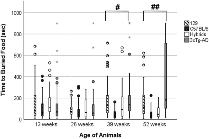
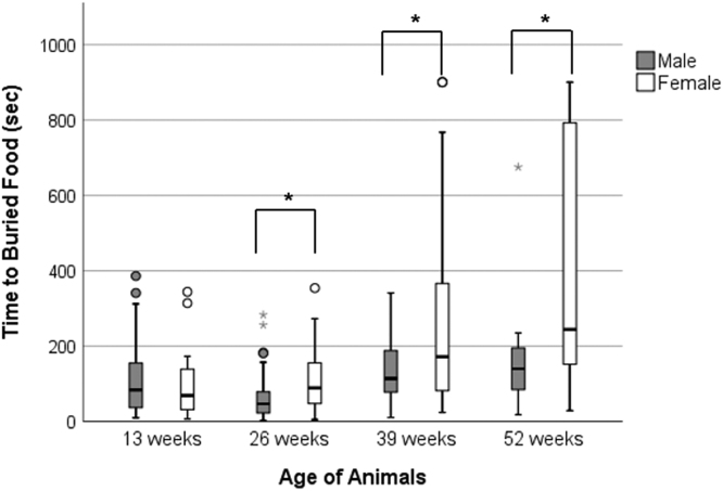
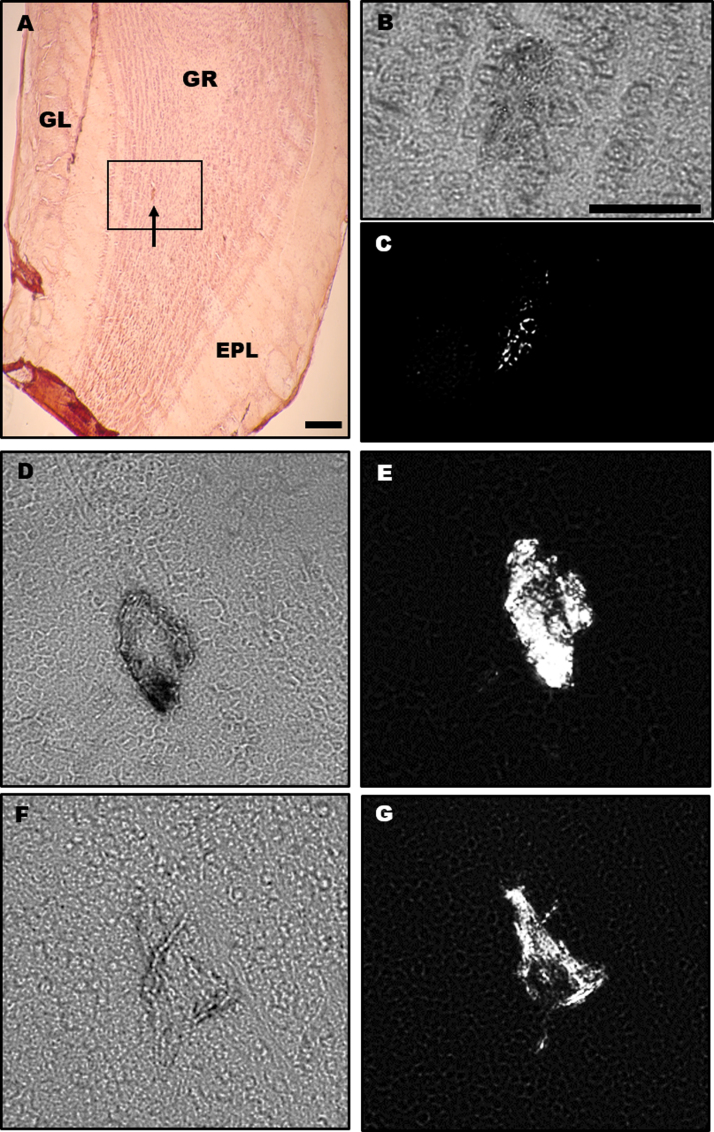
Alzheimer's disease (AD) is an incurable neurodegenerative disease in which the risk of development increases with age. People with AD are plagued with deficits in their cognition, memory, and basic social skills. Many of these deficits are believed to be caused by the formation of amyloid-β plaques and neurofibrillary tangles in regions of the brain associated with memory, such as the hippocampus. However, one of the early, preclinical symptoms of AD is the loss of olfactory detection and discrimination. To determine if a mouse model of AD expresses the same olfactory dysfunction seen in human AD, 3xTg-AD mice were given a buried food test and, unlike previous studies, compared to their background and parental strains. Results showed that over 52 weeks, the 3xTg-AD mice took significantly longer to find the buried food than the control strains. The olfactory bulbs of the 3xTg-AD mice were removed, sliced, and stained using Congo red for histological analysis. Amyloid deposits were observed predominantly in the granule layer of the olfactory bulb beginning at 13 weeks of age in 3xTg-AD mice, but not in the control strains of mice. Further examination of the buried food test data revealed that 3xTg-AD females had a significantly longer latency to detect the buried food than males beginning at 26 weeks of age. Overall, this study provides further validation of the 3xTg-AD mouse model of AD and supports the idea that simple olfactory testing could be part of the diagnostic process for human AD.
Keywords: 3xTg-AD; Alzheimer’s; Buried food test; Congo red; Olfaction.
© 2021 The Authors.
Conflict of interest statement
The authors have no conflicts of interest to report.
Figures
References
-
- Adlimoghaddam A., Snow W.M., Stortz G., Perez C., Djordjevic J., Goertzen A.L., Ko J.H., Albensi B.C. Regional hypometabolism in the 3xTg mouse model of Alzheimer’s disease. Neurobiol. Dis. 2019;127:264–277. - PubMed
-
- Aizenstein H.J., Nebes R.D., Saxton J.A., Price J.C., Mathis C.A., Tsopelas N.D., Ziolko S.K., James J.A., Snitz B.E., Houck P.R., Bi W., Cohen A.D., Lopresti B.J., DeKosky S.T., Halligan E.M., Klunk W.E. Frequent amyloid deposition without significant cognitive impairment among the elderly. Arch. Neurol. 2008;65:1509–1517. - PMC - PubMed
-
- Alberts J.R., Galef B.G. Acute anosmia in the rat: a behavioral test of a peripherally-induced olfactory deficit. Physiol. Behav. 1971;6:619–621. - PubMed
-
- Alzheimer’s Association 2020 Alzheimer’s disease facts and figures. Alzheimer’s Dement. 2020;16:391–460. (+)
-
- Attems J., Walker L., Jellinger K.A. Olfaction and aging: a mini-review. Gerontology. 2015;61:485–490. - PubMed
LinkOut - more resources
Full Text Sources
Other Literature Sources
Molecular Biology Databases