. 2021 Mar 29:(169):10.3791/62375.
doi: 10.3791/62375.

Dissection of the Endolymphatic Sac from Mice

Affiliations

Dissection of the Endolymphatic Sac from Mice

Keiji Honda et al. J Vis Exp. .

Abstract

The study of mutant mouse models of human hearing and balance disorders has unraveled many structural and functional changes which may contribute to the human phenotypes. Although important progress has been done in the understanding of the development and function of the neurosensory epithelia of the cochlea and vestibula, limited knowledge is available regarding the development, cellular composition, molecular pathways and functional characteristics of the endolymphatic sac. This is, in large part, due to the difficulty of visualizing and microdissecting this tissue, which is an epithelium comprised of only one cell layer. The study presented here describes an approach to access and microdissect the endolymphatic sac from the wild-type mouse inner ear at different ages. The result of a similar dissection is shown in a pendrin-deficient mouse model of enlargement of the vestibular aqueduct. A transgenic mouse with a fluorescent endolymphatic sac is presented. This reporter mouse can be used to readily visualize the endolymphatic sac with limited dissection and determine its size. It can also be used as an educational tool to teach how to dissect the endolymphatic sac. These dissection procedures should facilitate further characterization of this understudied part of the inner ear.


Figures

Figure 1.
Figure 1.. Whole-mount dissection of the endolymphatic sac of a wild type mouse at post natal day 5 (P5).
(A) Schematic of the membranous labyrinth of a developed mouse inner ear (right). The location of the endolymphatic sac, and the cochlea and vestibular structures are indicated. This figure has been modified from Honda et al.. (B) Mid-sagittal section showing the left hemi-skull. The location of the endolymphatic sac is indicated (arrow). (O, occipital; F, frontal) (C) Otic capsule along with the squamous part of the temporal bone. (D) Important landmarks overlaid onto the dotted box in panel C. (ES, endolymphatic sac; ED, endolymphatic duct; VA, vestibular aqueduct; ASC, anterior semicircular canal; PSC, posterior semicircular canal; CC, common crus; SS, sigmoid sinus) (E) Schematic illustration of the cross-section along with the continuous white line on panel D through the endolymphatic sac. The proximal side of the endolymphatic sac is covered by a bony canal called the vestibular aqueduct, along with the endolymphatic duct. In the distal side, the endolymphatic sac, surrounded by conjonctive tissue (CT), extends and protrudes to the outside of the bony labyrinth and is sandwiched between the dura mater (DM) and sigmoid sinus with the squamous part of the temporal bone (TB). (F) A fine needle is used to cut around the endolymphatic sac (following the dotted yellow lines on panel D). (G) The preparation is carefully removed from the temporal bone by holding the tissue at the position shown by the asterisk (*, also shown in panel E) and peeling it up. (H) An isolated whole endolymphatic sac with surrounding tissues. (I) Schematic version of this isolated endolymphatic sac with surrounding tissues. (J) Isolated endolymphatic sac without surrounding tissues. Scale bars: 22 mm (B), 1 mm (J). Please click here to view a larger version of this figure.
Figure 2.
Figure 2.. Dissection of the opened endolymphatic sac of a P5 wild type mouse.
(A) On step 2.4, the incision line on the vestibular aqueduct is positioned slightly anterior (red dotted line) to make an incision into the endolymphatic sac lumen. (B) Hold the stem part of the preparation, insert a 27-gauge needle into the lumen, and move it to cut the endolymphatic sac into two sheets. (C) Schematic illustration of panel B. (D) Hold the edge of each sheet with forceps and separate them from each other. (E) Schematic illustration of panel D. (F) The endolymphatic sac is separated into two sheets, including the epithelium and the surrounding tissues. Please click here to view a larger version of this figure.
Figure 3.
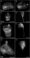
Figure 3.. Dissection of R26LSL-RCL-tdT/+;Tg(ATP6V1B1-Cre)1Rnel/Mn mouse endolymphatic sac at E16.5, P5, and P30.
All images presented show tdTomato fluorescence obtained using a stereomicroscope with a 1 x objective equipped for detection of tdTomato fluorescence. (A, B) Mid-sagittal section of the skull of a E16.5 R26LSL-RCL-tdT/+ ;Tg(ATP6V1B1-Cre)1Rnel/Mn mouse before (A) and after (B) half-brain removal. tdTomato fluorescence outlines the position of the inner ear. The endolymphatic sac is readily visible even without dissection (arrowhead). (C, E, G) Isolated inner ears from E16.5, P5 and P30 mice, respectively. (D, F and H) Higher magnification images of the corresponding microdissected endolymphatic sacs and ducts. At P30, the endolymphatic duct is encapsulated in bone making it particularly difficult to isolate. Scale bars: 2 mm (A, B, C, E, G), 500 μm (D, F, H). Please click here to view a larger version of this figure.
Figure 4.
Figure 4.. Gross anatomy of the otic capsule with an enlarged endolymphatic sac.
Inner ears from Slc26a4+/− (left) and Slc26a4−/− (right) mouse littermates at P105. The vestibular aqueduct and endolymphatic sac and duct (black dotted lines) are enlarged in the Slc26a4−/− mouse as compared to those in a Slc26a4+/− mouse. Scale bar: 2 mm. Please click here to view a larger version of this figure.
Figure 5.
Figure 5.. SLC26A4 expression of mitochondria-rich cells in the endolymphatic sac epithelium at E16.5 and P5.
(A, B) Isolated endolymphatic sac from E16.5 mouse labeled with anti-SLC26A4 antibody (green) and phalloidin which labels β-actin (ACTB, red). (C) A low-magnification image of opened endolymphatic sac from P5 mouse labeled with an anti-SLC26A4 antibody (green). Phalloidin (ACTB, red) can be used to highlight the presence of the endolymphatic sac as well as the conjunctive tissue around it. (D) Isolated endolymphatic sac labeled with an anti-SLC26A4 antibody (green). (E) Schematic illustration of endolymphatic sac epithelium highlighting the presence of two cell types: the mitochondria-rich cells, which apical surface is covered with microvilli, and the ribosome-rich cells. (F-G) High-magnification images of endolymphatic sac epithelium at P5, after the endolymphatic sac was opened and labeled with an anti-SLC26A4 antibody (green) and phalloidin (ACTB, red). A representative image at the apical membrane level is shown in G, and a reconstructed cross-section from z-stack at the level of the punctate white line is shown in F. The nucleus of the cells is labeled with DAPI (blue). Scale bars: 100 μm (A, B); 200 μm (C, D); 20 μm (F, G). Please click here to view a larger version of this figure.

Similar articles

Cited by

References

    1. Dror AA, Brownstein Z, Avraham KB Integration of human and mouse genetics reveals pendrin function in hearing and deafness. Cellular Physiology and Biochemistry. 28 (3), 535–544 (2011). - PMC - PubMed
    1. Reardon W, CF OM, Trembath R, Jan H, Phelps PD Enlarged vestibular aqueduct: a radiological marker of pendred syndrome, and mutation of the PDS gene. QJM. 93 (2), 99–104 (2000). - PubMed
    1. Griffith AJ, Wangemann P Hearing loss associated with enlargement of the vestibular aqueduct: mechanistic insights from clinical phenotypes, genotypes, and mouse models. Hearing Research. 281 (1-2), 11–17 (2011). - PMC - PubMed
    1. Gurkov R, Pyyko I, Zou J, Kentala E What is Meniere’s disease? A contemporary re-evaluation of endolymphatic hydrops. Journal of Neurology. 263 Suppl 1 S71–81 (2016). - PMC - PubMed
    1. Eckhard A et al. Water channel proteins in the inner ear and their link to hearing impairment and deafness. Molecular Aspects of Medicine. 33 (5-6), 612–637 (2012). - PubMed

Publication types

LinkOut - more resources