. 2021 Oct 22;144(9):2709-2721.
doi: 10.1093/brain/awab153.

Distinctive clinical presentation and pathogenic specificities of anti-AK5 encephalitis

Affiliations

Distinctive clinical presentation and pathogenic specificities of anti-AK5 encephalitis

Sergio Muñiz-Castrillo et al. Brain. .

Abstract

Limbic encephalitis with antibodies against adenylate kinase 5 (AK5) has been difficult to characterize because of its rarity. In this study, we identified 10 new cases and reviewed 16 previously reported patients, investigating clinical features, IgG subclasses, human leucocyte antigen and CSF proteomic profiles. Patients with anti-AK5 limbic encephalitis were mostly male (20/26, 76.9%) with a median age of 66 years (range 48-94). The predominant symptom was severe episodic amnesia in all patients, and this was frequently associated with depression (17/25, 68.0%). Weight loss, asthenia and anorexia were also highly characteristic, being present in 11/25 (44.0%) patients. Although epilepsy was always lacking at disease onset, seizures developed later in a subset of patients (4/25, 16.0%). All patients presented CSF abnormalities, such as pleocytosis (18/25, 72.0%), oligoclonal bands (18/25, 72.0%) and increased Tau (11/14, 78.6%). Temporal lobe hyperintensities were almost always present at disease onset (23/26, 88.5%), evolving nearly invariably towards severe atrophy in subsequent MRIs (17/19, 89.5%). This finding was in line with a poor response to immunotherapy, with only 5/25 (20.0%) patients responding. IgG1 was the predominant subclass, being the most frequently detected and the one with the highest titres in nine CSF-serum paired samples. A temporal biopsy from one of our new cases showed massive lymphocytic infiltrates dominated by both CD4+ and CT8+ T cells, intense granzyme B expression and abundant macrophages/microglia. Human leucocyte antigen (HLA) analysis in 11 patients showed a striking association with HLA-B*08:01 [7/11, 63.6%; odds ratio (OR) = 13.4, 95% confidence interval (CI): 3.8-47.4], C*07:01 (8/11, 72.7%; OR = 11.0, 95% CI: 2.9-42.5), DRB1*03:01 (8/11, 72.7%; OR = 14.4, 95% CI: 3.7-55.7), DQB1*02:01 (8/11, 72.7%; OR = 13.5, 95% CI: 3.5-52.0) and DQA1*05:01 (8/11, 72.7%; OR = 14.4, 95% CI: 3.7-55.7) alleles, which formed the extended haplotype B8-C7-DR3-DQ2 in 6/11 (54.5%) patients (OR = 16.5, 95% CI: 4.8-57.1). Finally, we compared the CSF proteomic profile of five anti-AK5 patients with that of 40 control subjects and 10 cases with other more common non-paraneoplastic limbic encephalitis (five with antibodies against leucine-rich glioma inactivated 1 and five against contactin-associated protein-like 2), as well as 10 cases with paraneoplastic neurological syndromes (five with antibodies against Yo and five against Ma2). These comparisons revealed 31 and seven significantly upregulated proteins in anti-AK5 limbic encephalitis, respectively mapping to apoptosis pathways and innate/adaptive immune responses. These findings suggest that the clinical manifestations of anti-AK5 limbic encephalitis result from a distinct T cell-mediated pathogenesis, with major cytotoxicity-induced apoptosis leading to a prompt and aggressive neuronal loss, likely explaining the poor prognosis and response to immunotherapy.

Keywords: adenylate kinase 5; human leucocyte antigen; limbic encephalitis; proteomics.


Figures

Figure 1
Figure 1
Typical neuroimaging findings in anti-AK5 limbic encephalitis. (A) Coronal FLAIR MRI showing bilateral, enlarged, hyperintense medial temporal lobes from Patient 10. (B) Axial T1 MRI with gadolinium from the same patient, showing right medial temporal contrast enhancement. (C and D) Severe hippocampal atrophy in Patient 8 involving both medial temporal lobes on coronal (C) and axial (D) FLAIR-MRI.
Figure 2
Figure 2
Histopathological characteristics of anti-AK5 limbic encephalitis. Right uncal biopsy from Patient 10 showing intense perivascular and parenchymal inflammatory infiltrates (A, haematoxylin and eosin staining, ×200), which was dominated by T cells that were abundant in both the perivascular space and the parenchyma (B, CD3 immunostaining, ×200). Conversely, B cells, though highly present in the perivascular space, were scattered in the parenchyma (C, CD20 immunostaining, ×200). The T-cell infiltrate included both CD4+ (D, ×200) and CD8+ T cells (E, ×200), which were especially abundant in the perivascular space but also present diffusely in the parenchyma. Cytotoxic CD8+ T cells expressing granzyme B (a protein stored in the granules of cytotoxic cells that induces apoptosis) were detected in perivascular and parenchymal infiltrates (F, ×200). Nodular clusters of inflammatory cells were detected in the parenchyma using haematoxylin and eosin staining (G, ×400) and CD8+ and CD4+ T-cell clusters were also observed (H and I, CD8 and CD4 immunostainings, respectively, ×400). Additionally, macrophages/microglia diffusely infiltrated the tissue, as shown by CD68 (J, ×100) and CD163 (K, ×100) immunostainings, the latter being associated with M2 differentiation related to an anti-inflammatory response including clearance of apoptotic bodies.
Figure 3
Figure 3
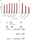
HLA analysis in patients with anti-AK5 limbic encephalitis. (A) Carrier frequencies (%) for the alleles of the ancestral haplotype 8.1 in the control group (n = 442) and anti-AK5 limbic encephalitis patients (n = 11). (B) Carrier frequencies (%) for different combined components of the ancestral haplotype 8.1 in the control group (n = 442) and patients with anti-AK5 limbic encephalitis (n = 11). (C) Location of AK5-derived peptides predicted as strong binders within the protein sequence. AK5-derived peptides predicted as strong HLA binders were located preferentially within the N-terminus of the protein, which is shared by the isoforms 1 and 3. The length of the dashed bar represents the amount of predicted peptides as strong binders for the HLA shown. Numbers correspond to the sequence of the canonical isoform 1.
Figure 4
Figure 4
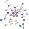
Network of upregulated CSF proteins in anti-AK5 limbic encephalitis compared to anti-LGI1/CASPR2 patients. The two more relevant pathways are highlighted: positive regulation of protein insertion into the mitochondrial membrane involved in apoptotic signalling pathway is coloured in red, while immune system process pathway is coloured in blue. Colour-coded lines indicate an association between two proteins: red, gene fusion; blue, gene co-occurrence; green, genome neighbourhood; black, co-expression; purple, biochemical or genetic experimental data; yellow, text mining; and light blue, known association in curated databases. C1R = complement 1; C4A = complement c4-a; C4B = complement c4-b; CA3 = carbonic anhydrase 3; CAPG = macrophage-capping protein; CD209 = CD 209 antigen; CD300C = CMRF35-like molecule 6; CSF3 = granulocyte colony-stimulating factor; FAM3B = protein FAM3B; GFAP = glial fibrillary acidic protein; GRN = progranulin; HAVCR2 = hepatitis A virus cellular receptor 2; ISG15 = ubiquitin-like protein 15; LYZ = lysozyme C; MB = myoglobin; MRC1 = macrophage mannose receptor 1; PEBP1 = phosphatidylethanolamine-binding protein 1; PGAM1 = phosphoglycerate mutase 1; PPIA = peptidyl-prolyl cis-trans isomerase A; PSME1 = proteasome activator complex subunit 1; S100A4 = protein S100-A4; SFN = protein 14-3-3 sigma; SLAMF7 = SLAM family member 7; UBE2I = SUMO-conjugating enzyme UBC9; UFC1 = ubiquitin-fold modifier-conjugation enzyme 1; YWHAB = protein 14-3-3 beta/alpha; YWHAE = protein 14-3-3 epsilon; YWHAG = protein 14-3-3 gamma; YWHAH = protein 14-3-3 eta; YWHAQ = protein 14-3-3 theta; YWHAZ = protein 14-3-3 zeta/delta.
Figure 5
Figure 5
Heat map of CSF expression of proteins involved in the most relevant biological pathways. CSF expression for proteins involved in the two most relevant biological pathways (apoptosis and immune system) is shown as the median Z-score transformed for every protein in each group. All proteins were significantly upexpressed in anti-AK5 patients (n = 5) compared to anti-CASPR2/LGI1 (n = 5 each) cases, and their expression was also higher when compared to paraneoplastic neurological syndromes with antibodies against Yo/Ma2 (n = 5 each), although only those marked with an asterisk were statistically significant. C1R = complement 1; C4A = complement c4-a; C4B = complement c4-b; CD209 = CD 209 antigen; CD300C = CMRF35-like molecule 6; CSF3 = granulocyte colony-stimulating factor; GRN = progranulin; HAVCR2 = hepatitis A virus cellular receptor 2; ISG15 = ubiquitin-like protein 15; LYZ = lysozyme C; MB = myoglobin; MRC1 = macrophage mannose receptor 1; PGAM1 = phosphoglycerate mutase 1; PPIA = peptidyl-prolyl cis-trans isomerase A; PSME1 = proteasome activator complex subunit 1; SFN = protein 14-3-3 sigma; SLAMF7 = SLAM family member 7; YWHAB = protein 14-3-3 beta/alpha; YWHAE = protein 14-3-3 epsilon; YWHAG = protein 14-3-3 gamma; YWHAH = protein 14-3-3 eta; YWHAQ = protein 14-3-3 theta; YWHAZ = protein 14-3-3 zeta/delta.

References

    1. Tüzün E, Rossi JE, Karner SF, Centurion AF, Dalmau J.. Adenylate kinase 5 autoimmunity in treatment refractory limbic encephalitis. J Neuroimmunol. 2007;186(1-2):177–180. - PMC - PubMed
    1. Do L-D, Chanson E, Desestret V, et al.Characteristics in limbic encephalitis with anti–adenylate kinase 5 autoantibodies. Neurology. 2017;88(6):514–524. - PubMed
    1. Bien CI, Nehls F, Kollmar R, et al.Identification of adenylate kinase 5 antibodies during routine diagnostics in a tissue-based assay: Three new cases and a review of the literature. J Neuroimmunol. 2019;334:576975. - PubMed
    1. Zoccarato M, Valeggia S, Zuliani L, et al.Conventional brain MRI features distinguishing limbic encephalitis from mesial temporal glioma. Neuroradiology. 2019;61(8):853–860. - PubMed
    1. Vogrig A, Joubert B, André‐Obadia N, Gigli GL, Rheims S, Honnorat J.. Seizure specificities in patients with antibody‐mediated autoimmune encephalitis. Epilepsia. 2019;60(8):1508–1525. - PubMed

Publication types