. 2021 May 28;41(5):BSR20210198.
doi: 10.1042/BSR20210198.

Paracrine interleukin-8 affects mesenchymal stem cells through the Akt pathway and enhances human umbilical vein endothelial cell proliferation and migration

Affiliations

Paracrine interleukin-8 affects mesenchymal stem cells through the Akt pathway and enhances human umbilical vein endothelial cell proliferation and migration

Lulu Wang et al. Biosci Rep. .

Retraction in

Abstract

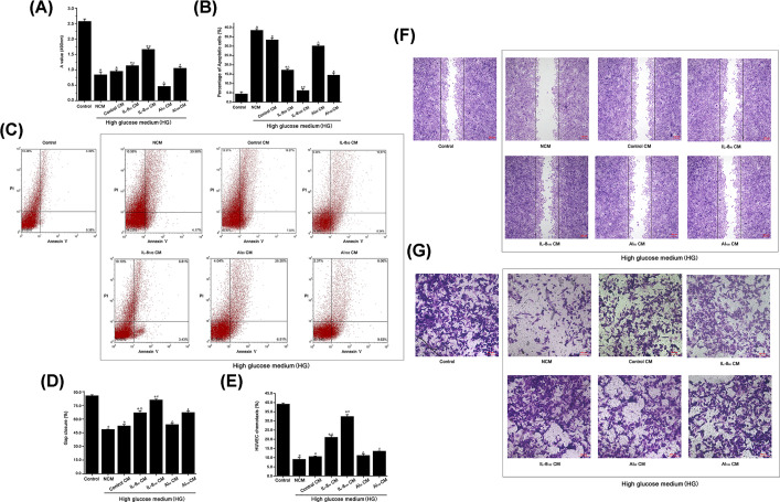
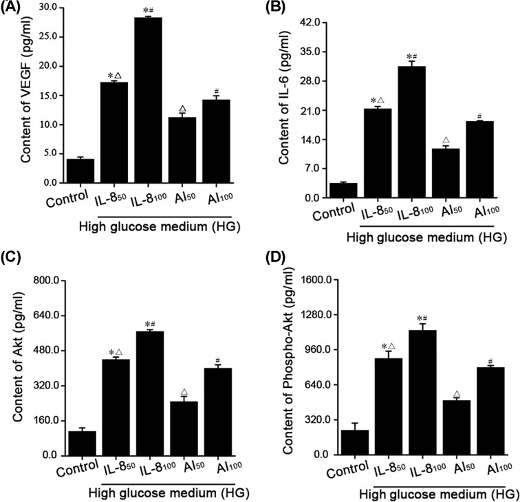
Interleukin-8 (IL-8) promotes cell homing and angiogenesis, but its effects on activating human bone marrow mesenchymal stem cells (BMSCs) and promoting angiogenesis are unclear. We used bioinformatics to predict these processes. In vitro, BMSCs were stimulated in a high-glucose (HG) environment with 50 or 100 μg/ml IL-8 was used as the IL-8 group. A total of 5 μmol/l Triciribine was added to the two IL-8 groups as the Akt inhibitor group. Cultured human umbilical vein endothelial cells (HUVECs) were cultured in BMSCs conditioned medium (CM). The changes in proliferation, apoptosis, migration ability and levels of VEGF and IL-6 in HUVECs were observed in each group. Seventy processes and 26 pathways were involved in vascular development, through which IL-8 affected BMSCs. Compared with the HG control group, HUVEC proliferation absorbance value (A value), Gap closure rate, and Transwell cell migration rate in the IL-8 50 and IL-8 100 CM groups were significantly increased (P<0.01, n=30). However, HUVEC apoptosis was significantly decreased (P<0.01, n=30). Akt and phospho-Akt (P-Akt) protein contents in lysates of BMSCs treated with IL-8, as well as VEGF and IL-6 protein contents in the supernatant of BMSCs treated with IL-8, were all highly expressed (P<0.01, n=15). These analyses confirmed that IL-8 promoted the expression of 41 core proteins in BMSCs through the PI3K Akt pathway, which could promote the proliferation and migration of vascular endothelial cells. Therefore, in an HG environment, IL-8 activated the Akt signaling pathway, promoted paracrine mechanisms of BMSCs, and improved the proliferation and migration of HUVECs.

Keywords: Bioinformatics; Human bone marrow mesenchymal stem cells; Human umbilical vein endothelial cells; Interleukin-8; Migration; Proliferation.


Conflict of interest statement

The authors declare that there are no competing interests associated with the manuscript.

Figures

Figure 1
Figure 1. Gene expression of IL-8 and BMSC
(A) Venn analysis of IL-8 and BMSC gene expression. (B) Common gene expression profile heat map of IL-8 and BMSCs as detected by the GSE9520 dataset. The Pearson correlation distance metric and the average linkage clustering algorithm were used.
Figure 2
Figure 2. GO analysis of the genes common to IL-8 and BMSCs
(A–C) Enrichment analysis histogram of the BP, CC, and MF of the genes common to IL-8 and BMSCs. The y-axis indicates different GO terms and the x-axis indicates the enrichment score in each category. (D–F) Chordal graph of IL-8 and BMSCs common genes with BP, CC, and MF.
Figure 3
Figure 3. Common genes pathway analysis of IL-8 and BMSCs
(A) Pathway enrichment analysis bubble chart of genes common to IL-8 and BMSC. The y-axis indicates different pathway terms and the x-axis indicates the enrichment score in each category. (B) Chordal graph of genes common to IL-8 and BMSCs with pathways.
Figure 4
Figure 4. Network construction of IL-8 and BMSC gene protein interaction, and screening of core proteins
(A) The protein interaction network of IL-8 and BMSC genes. (B) Strategy diagram of IL-8 and BMSC core protein screening. The node area or font size is positively correlated with the wiring of the node. DC, BC, Closeness centrality (CC), NC, LAC.
Figure 5
Figure 5. In an HG environment, IL-8 stimulated BMSC-CM increased HUVEC proliferation and migration, as well as inhibited HUVEC apoptosis
(A) MTT assay was used to detect the proliferation of HUVECs, *P<0.05, #P<0.01, P<0.01 (n=30). (B) The comparison of HUVEC apoptosis rate in each group, *P<0.01, #P<0.01, P<0.01 (n=30). (C) HUVEC apoptosis was detected by Annexin V-PI flow cytometry. (D) Scratch closure rate of HUVECs in each group. *P<0.05, #P<0.05, P<0.05 (n=30). (E) The migration rate of HUVECs in each group. *P<0.01, #P<0.01, P<0.01 (n=30). (F) Representative images of wound healing in each treatment group after 24 h of culture. Scale bar = 100 µm. The black line represents the scratch area at 0 h. (G) Representative images of HUVEC migration in each treatment group after 24 h of culture. Scale bar = 100 µm.
Figure 6
Figure 6. IL-8 promoted Akt protein expression in HG-IL-850 and HG-IL-8100 groups, as well as up-regulation of VEGF and IL-6 protein
(A) The content of VEGF protein in the supernatant of BMSCs was determined by ELISA. *P<0.01, #P<0.05, P<0.01 (n=15). (B) The content of IL-6 protein in each BMSC supernatant was determined by ELISA. *P<0.01, #P<0.01, P<0.01 (n=15). (C) The content of Akt protein in BMSC lysates was determined by ELISA. *P<0.01, #P<0.01, P<0.01 (n=15). (D) The content of P-Akt protein in each BMSC cohort was determined by ELISA. *P<0.01, #P<0.01, P<0.01 (n=15).

Similar articles

Cited by

References

    1. Saeedi P., Petersohn I., Salpea P.et al. . (2019) Global and regional diabetes prevalence estimates for 2019 and projections for 2030 and 2045: results from the International Diabetes Federation Diabetes Atlas, 9th edition. Diabetes Res. Clin. Pract. 157, 107843 10.1016/j.diabres.2019.107843 - DOI - PubMed
    1. Chan J.C., Lim L.L., Wareham N.J.et al. . (2020) The Lancet Commission on diabetes: using data to transform diabetes care and patient lives. Lancet 396, 2019–2082 10.1016/S0140-6736(20)32374-6 - DOI - PubMed
    1. Eming S.A., Martin P. and Tomic-Canic M. (2014) Wound repair and regeneration: mechanisms, signaling, and translation. Sci. Transl. Med. 6, 265sr6 10.1126/scitranslmed.3009337 - DOI - PMC - PubMed
    1. Nigi L., Fondelli C., Donato G., Palasciano G., Setacci C. and Dotta F. (2018) Fighting diabetic foot ulcers-The diabetologist: A king maker of the fight. Semin. Vasc. Surg. 31, 49–55 10.1053/j.semvascsurg.2018.12.003 - DOI - PubMed
    1. Schleier L., Wiendl M., Heidbreder K.et al. . (2020) Non-classical monocyte homing to the gut via α4β7 integrin mediates macrophage-dependent intestinal wound healing. Gut 69, 252–263 10.1136/gutjnl-2018-316772 - DOI - PubMed

Publication types