. 2021 Apr 12;116(1):24.
doi: 10.1007/s00395-021-00866-8.

Omecamtiv mecarbil evokes diastolic dysfunction and leads to periodic electromechanical alternans

Affiliations

Omecamtiv mecarbil evokes diastolic dysfunction and leads to periodic electromechanical alternans

Gábor Á Fülöp et al. Basic Res Cardiol. .

Abstract

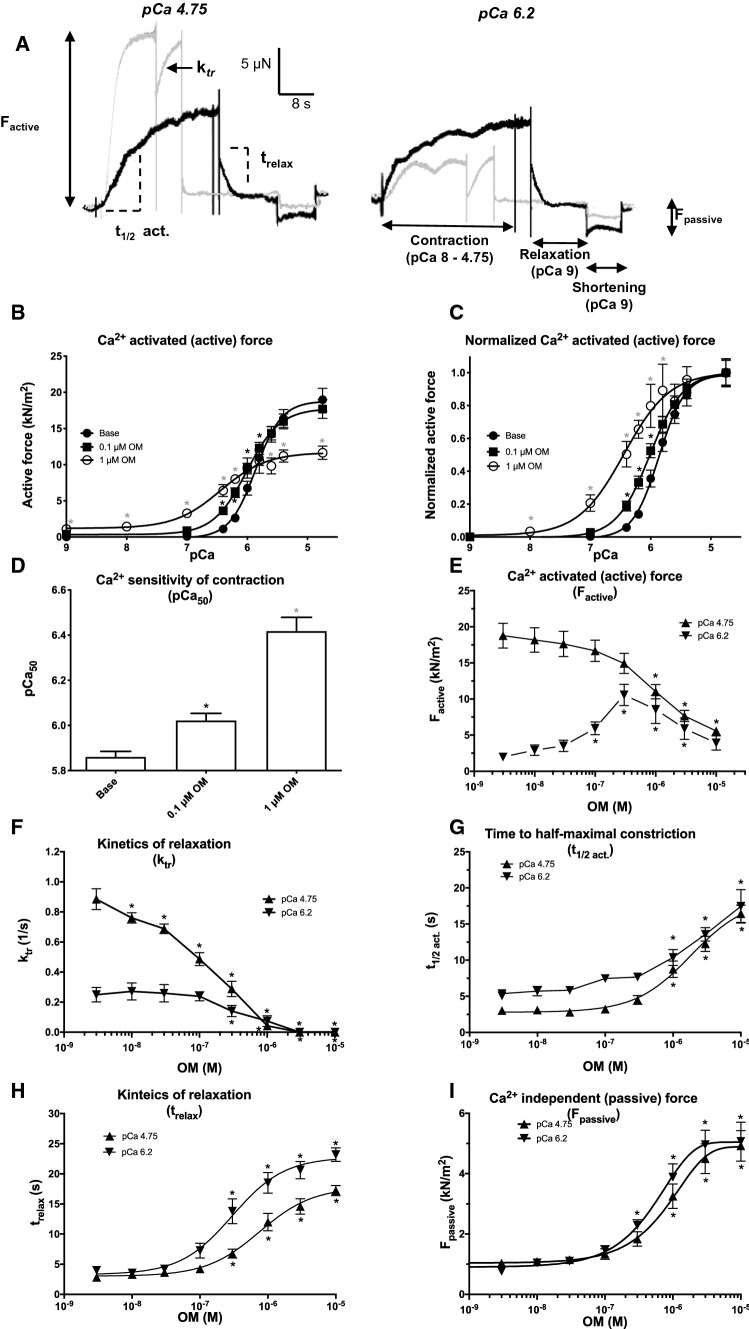
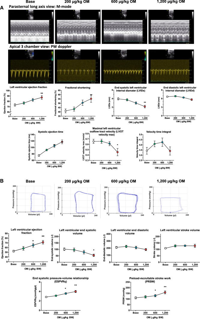
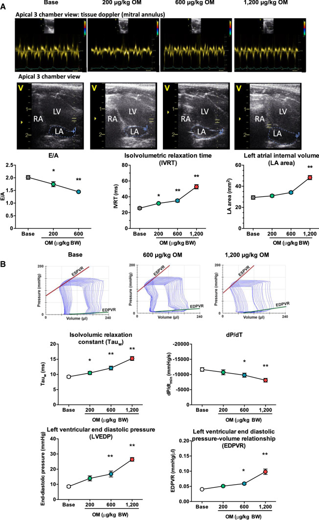
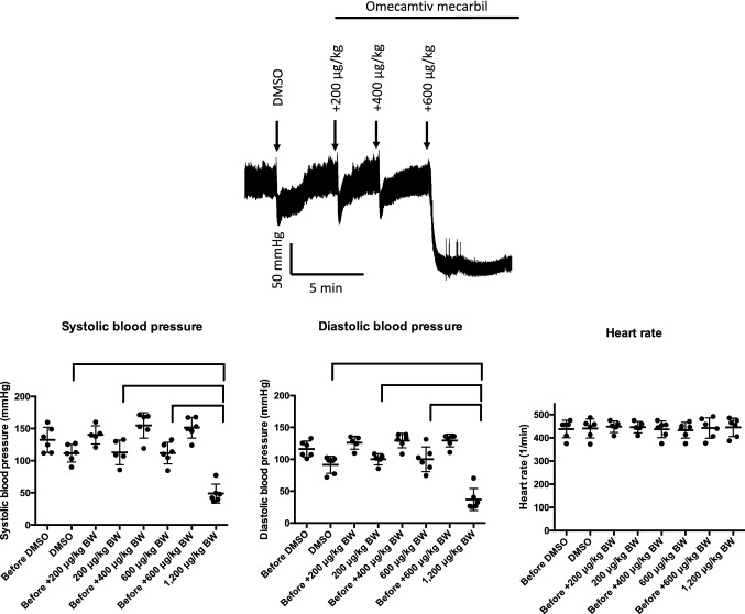
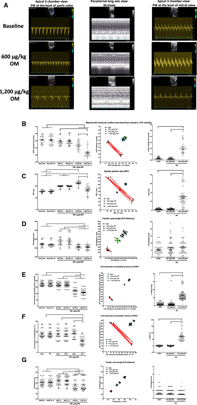
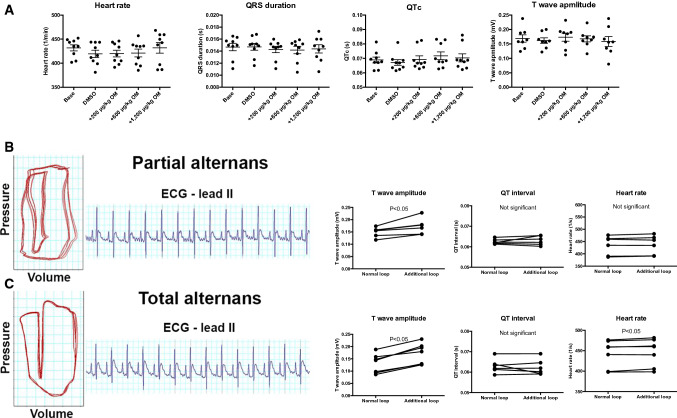
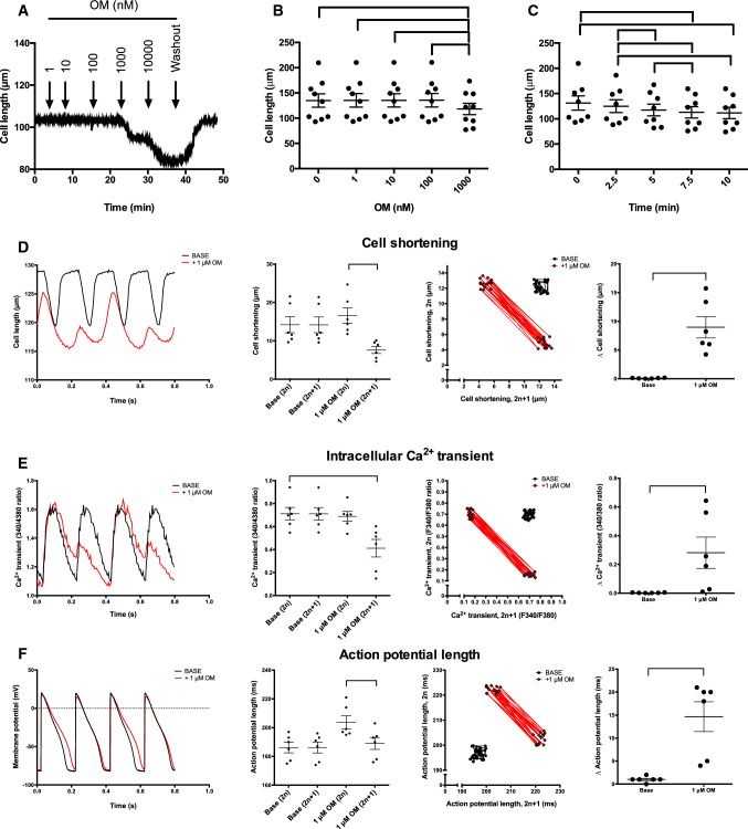
Omecamtiv mecarbil (OM) is a promising novel drug for improving cardiac contractility. We tested the therapeutic range of OM and identified previously unrecognized side effects. The Ca2+ sensitivity of isometric force production (pCa50) and force at low Ca2+ levels increased with OM concentration in human permeabilized cardiomyocytes. OM (1 µM) slowed the kinetics of contractions and relaxations and evoked an oscillation between normal and reduced intracellular Ca2+ transients, action potential lengths and contractions in isolated canine cardiomyocytes. Echocardiographic studies and left ventricular pressure-volume analyses demonstrated concentration-dependent improvements in cardiac systolic function at OM concentrations of 600-1200 µg/kg in rats. Administration of OM at a concentration of 1200 µg/kg was associated with hypotension, while doses of 600-1200 µg/kg were associated with the following aspects of diastolic dysfunction: decreases in E/A ratio and the maximal rate of diastolic pressure decrement (dP/dtmin) and increases in isovolumic relaxation time, left atrial diameter, the isovolumic relaxation constant Tau, left ventricular end-diastolic pressure and the slope of the end-diastolic pressure-volume relationship. Moreover, OM 1200 µg/kg frequently evoked transient electromechanical alternans in the rat in vivo in which normal systoles were followed by smaller contractions (and T-wave amplitudes) without major differences on the QRS complexes. Besides improving systolic function, OM evoked diastolic dysfunction and pulsus alternans. The narrow therapeutic window for OM may necessitate the monitoring of additional clinical safety parameters in clinical application.

Keywords: Ca2+ sensitivity; Diastolic dysfunction; Heart failure; Inotropy; Omecamtiv mecarbil; Pulsus alternans.


Conflict of interest statement

The authors have declared that no conflicts of interest exist.

Figures

Fig. 1
Fig. 1
Omecamtiv mecarbil evokes Ca2+ sensitization, slower contraction–relaxation kinetics and increased passive stiffness in permeabilized human cardiomyocytes. Original force recordings at maximal (pCa 4.75; ≈18 μM) and submaximal (pCa 6.2; ≈0.63 μM) Ca2+ concentrations illustrate the effects of 1 µM omecamtiv mecarbil (OM) on force development in isolated, permeabilized human left ventricular cardiomyocytes. Gray traces: controls; black traces: the same cells with OM (a). Steps of the measurement are indicated at pCa 6.2 (black trace). First, the permeabilized ventricular cardiomyocyte was placed in a Ca2+-containing solution and the developing Ca2+-dependent force was recorded. Then the preparation was transferred to a relaxing solution (pCa 9). After steady-state relaxation, the passive stiffness was determined by shortening the sarcomeres to a slack level. The Ca2+ concentration–active force relationship was determined (shown in absolute and relative [normalized] units in panels (b) and (c), respectively) without (baseline) and with 0.1 and 1 µM OM (as indicated). The bar graph (d) represents the mean ± standard error of the mean (SEM) of seven to nine individual measurements (a significant difference [P < 0.05] from baseline is indicated by an asterisk). OM had a biphasic effect on the active force development as shown by the concentration–response graph at pCa 4.75 (e). The overall rate of active force development (ktr; f) and the time required for half-maximal Ca2+-dependent force development (t1/2 act; g) are shown on the graphs. Finally, the rate of relaxation (trelax; H) and the passive (Ca2+-independent) stiffness (Fpassive; i) were also determined at different OM concentrations. The applied OM concentrations are shown on the horizontal axes. Symbols represent the mean of seven to nine independent determinations. Error bars indicate the SEM
Fig. 2
Fig. 2
Omecamtiv mecarbil improves left ventricular systolic function in the rat. Omecamtiv mecarbil (OM) was tested in the rat in vivo at cumulative doses of 200–1200 µg/kg body weight. Left ventricular systolic function was studied by echocardiography (a) and by left ventricular pressure–volume analysis (b). M-mode was used in the parasternal long-axis view (representative recordings shown in the upper row) to determine ejection fraction, fractional shortening, left ventricular end-systolic internal diameter and left ventricular end-diastolic internal diameter. The pulsatile wave (PW) Doppler method (from the left ventricular outflow tract) was used to determine hemodynamic parameters (representative recordings in the lower row of traces), such as left ventricular systolic ejection time, maximal blood flow velocity at the left ventricular outflow tract and left ventricular outflow tract velocity time integral, as shown in the graphs. Representative left ventricular steady-state pressure–volume loops were obtained at different cumulative OM doses during pressure–volume analysis (b). Ejection fraction, end-systolic volume, end-diastolic volume, stroke volume and load-independent contractility indices (slope of the end-systolic pressure–volume relationship; preload recruitable stroke work) at different OM doses are shown in the graphs. The number of independent observations was eight to 14 for echocardiography and nine for the pressure–volume analysis. Symbols represent the mean and standard error of the mean. Significant differences from the initial (baseline) values upon application of OM (cumulative doses are shown on the horizontal axes) are indicated by asterisks: *P < 0.5; **P < 0.01
Fig. 3
Fig. 3
Omecamtiv mecarbil negatively affects left ventricular diastolic filling in the rat. Omecamtiv mecarbil (OM) was tested in the rat in vivo. Left ventricular diastolic function was studied by echocardiography (a) and by left ventricular pressure–volume analysis (b). Pulsatile-wave Doppler inflow at the mitral valve was used to determine the early:atrial filling ratio (E:A). The tissue Doppler method at the mitral annulus (representative individual recordings in the top row) was used to determine isovolumetric relaxation time. Left atrial area was determined from two-dimensional images in an apical 3 chamber view (representative pictograms are shown in the row below the tissue Doppler recordings) and values are plotted on the graphs. Representative original recordings of left ventricular pressure–volume relationships during transient occlusion of the inferior vena cava at different OM doses are shown in the top row of panel (b). The following diastolic parameters were acquired from pressure–volume analysis: indices of left ventricular active relaxation (dP/dtmin and the isovolumic relaxation constant) and left ventricular stiffness (end-diastolic pressure and slope of the end-diastolic pressure–volume relationship). The number of independent observations was seven to 14 for echocardiography and nine for the pressure–volume analysis. Symbols represent the mean and standard error of the mean. Significant differences from the initial (baseline) values upon application of OM (cumulative doses are shown on the horizontal axes) are indicated by asterisks: *P < 0.5; **P < 0.01
Fig. 4
Fig. 4
Omecamtiv mecarbil evokes severe hypotension in the rat. Omecamtiv mecarbil (OM) was tested in the rat in vivo. Application of solvent (dimethyl sulfoxide [DMSO]) alone and increasing doses of OM are shown in a representative invasive blood pressure recording at the top. Systolic and diastolic blood pressure and heart rate were determined immediately before and 5 min after bolus injections as indicated on the horizontal axes. Each symbol represents an individual measurement. The mean and standard error of the measurements are shown on the scatter graphs. Significant (P < 0.05) differences among the six replicates are indicated by the braces
Fig. 5
Fig. 5
Omecamtiv mecarbil evokes transient pulsus alternans in the rat. High doses (1200 µg/kg body weight cumulative dose) of omecamtiv mecarbil (OM) evoked a transient (short-lasting, recurring) electromechanical alternans (alternating pulseless electrical activity) in the rat. It was observed during left ventricular (LV) pressure–volume analysis (a) and invasive blood pressure measurements (bd). The features and development of the electromechanical alternans are shown by representative experiments. An individual, representative recording of the parallel measurement of LV pressure, volume, heart rate (HR) and the electrocardiogram (ECG) are shown in the upper row of traces in panel (a). Features at baseline and on the development of partial and total alternans are shown in the second row of traces in panel (a), which illustrate the raw pressure and volume values and the loops. The development of the alternans is also shown in representative recordings by means of invasive arterial blood pressure measurements (b, c). The electromechanical alternans developed upon a slight increase in heart rate (a, b) or upon an extrasystole (c). Irrespective of its initiation, alternans was characterized by pulseless electrical activity that repeatedly appeared after an normal beat, as represented by the parallel-pressure waveform and ECG recordings at baseline and with a cumulative OM dose of 1200 µg/kg (d). Single, representative recordings are shown and were specifically chosen to illustrate this feature
Fig. 6
Fig. 6
Echocardiographic features of omecamtiv mecarbil evoked transient, periodic electromechanical alternans in the rat. Echocardiography (ad) and left ventricular pressure–volume relationships (eg) were recorded to evaluate the electromechanical alternans evoked by a 1200 µg/kg body weight (BW) dose of omecamtiv mecarbil (OM). Representative echocardiographic recordings are shown in panel (a). Each symbol represents an individual measurement (12 consecutive cardiac cycles in each animal), together with the mean and standard error, in the graphs on the left-hand side. The beat-to-beat variability is shown on the Poincaré plots in the middle set of graphs, where the value for the actual beat is plotted as the function of the value at the next beat in an individual representative recording. On the Poincaré plots, consecutive values are connected by lines. The cumulative dose of OM is indicated on the horizontal axes or in the insets (Poincaré plots). The difference between the consecutive beat pairs is plotted in the graphs on the right-hand side. Significant differences (P < 0.05) between the five to six (echocardiography) and nine (pressure–volume analysis) biological replicates are indicated by the braces. Left ventricular outflow tract velocity (b) and left ventricular systolic ejection time (c) were determined by pulsatile Doppler at the level of the aortic valve in an apical 3 chamber view, while the cardiac cycle length (d) was determined by a parallel electrocardiography recording. The characteristics of the electromechanical alternans were also recorded by means of left ventricular pressure–volume relationships, with data plotted similarly to the echocardiography data (eg). Left ventricular end-systolic pressure (e) and left ventricular end-diastolic volume (f) were determined by means of a probe inserted into the left ventricle, whereas the cardiac cycle length (g) was determined from the parallel ECG recording
Fig. 7
Fig. 7
Omecamtiv mecarbil evokes a transient T-wave alternans in the rat. Cardiac electrocardiograms were recorded in rats. A representative tracing and pooled analysis are shown. Heart rate, QRS duration, corrected QT interval (QTc; Bazett formula) and T-wave amplitude were recorded and plotted in periods without electromechanical alternans in the presence of 1200 µg/kg body weight omecamtiv mecarbil (OM) (a). Symbols represent individual values from eight to nine replicates. The mean and standard error of the mean are shown in the scatter plots. No statistical differences were found between the groups. Next, cardiac electrocardiography (ECG) parameters were evaluated during the transient periods when partial or total electromechanical alternans was present (b). Representative pressure–volume loops and ECG recordings are shown on the left-hand side. The graphs on the right-hand side represent the T-wave amplitudes, QT interval and heart rate values in the normal and consecutive diminished (additional) loops. Symbols represent the individual values determined from six biological replicates. Significant differences are indicated in the graphs
Fig. 8
Fig. 8
Omecamtiv mecarbil affects Ca2+ cycling in isolated intact canine left ventricular cardiomyocytes. Representative recordings are shown in panels (ac). The cell lengths were evaluated after 10 min of incubation with the indicated concentrations (0–1000 nM; n = 9; b) of omecamtiv mecarbil (OM). The kinetics of 1 μM OM evoked unloaded (occurring without stimulation) reduction of diastolic length of cardiomyocytes, as shown in panel (c). Each symbol represents a measurement on an individual cell. The mean is represented by horizontal lines and the error bars represent the standard error of the mean. In the second set of experiments, the cell length (optical measurement; d), intracellular Ca2+ concentration (measurement of changes in FURA-2 fluorescence intensity ratios; e) and membrane potential (measured by patch clamp; f) were measured in parallel on the same isolated cardiomyocyte. The stimulation frequency was 5 Hz. The recordings before OM treatment are labeled ‘BASE’, while the traces recorded from the same cell in the presence of OM are labeled ‘ + 1 μM OM’. Representative recordings are shown in the graphs on the left-hand side. Six biological replicates were recorded for all parameters and evaluated. Each symbol represents a single measurement on an individual cell. The mean is represented by horizontal lines and the error bars represent the standard error of the mean. To visualize the variations in consecutive values, Poincaré plots were constructed involving 30 consecutive cycles. In these plots, the parameter values of the even-numbered cycles (2n) are plotted as a function of the value of the next odd-numbered cycle (2n + 1). Finally, in the right-hand graphs, each symbol represents the average variability of an individual cell. Significant differences (P < 0.05) between groups are shown by the braces

References

    1. Bakkehaug JP, Kildal AB, Engstad ET, Boardman N, Naesheim T, Ronning L, Aasum E, Larsen TS, Myrmel T, How OJ. Myosin activator omecamtiv mecarbil increases myocardial oxygen consumption and impairs cardiac efficiency mediated by resting myosin ATPase activity. Circ Heart Fail. 2015;8:766–775. doi: 10.1161/CIRCHEARTFAILURE.114.002152. - DOI - PubMed
    1. Baudenbacher F, Schober T, Pinto JR, Sidorov VY, Hilliard F, Solaro RJ, Potter JD, Knollmann BC. Myofilament Ca2+ sensitization causes susceptibility to cardiac arrhythmia in mice. J Clin Invest. 2008;118:3893–3903. doi: 10.1172/JCI36642. - DOI - PMC - PubMed
    1. Cleland JG, Teerlink JR, Senior R, Nifontov EM, Mc Murray JJ, Lang CC, Tsyrlin VA, Greenberg BH, Mayet J, Francis DP, Shaburishvili T, Monaghan M, Saltzberg M, Neyses L, Wasserman SM, Lee JH, Saikali KG, Clarke CP, Goldman JH, Wolff AA, Malik FI. The effects of the cardiac myosin activator, omecamtiv mecarbil, on cardiac function in systolic heart failure: a double-blind, placebo-controlled, crossover, dose-ranging phase 2 trial. Lancet. 2011;378:676–683. doi: 10.1016/S0140-6736(11)61126-4. - DOI - PubMed
    1. Diaz ME, Eisner DA, O'Neill SC. Depressed ryanodine receptor activity increases variability and duration of the systolic Ca2+ transient in rat ventricular myocytes. Circ Res. 2002;91:585–593. doi: 10.1161/01.res.0000035527.53514.c2. - DOI - PubMed
    1. Greenberg BH, Chou W, Saikali KG, Escandon R, Lee JH, Chen MM, Treshkur T, Megreladze I, Wasserman SM, Eisenberg P, Malik FI, Wolff AA, Shaburishvili T. Safety and tolerability of omecamtiv mecarbil during exercise in patients with ischemic cardiomyopathy and angina. JACC Heart Fail. 2015;3:22–29. doi: 10.1016/j.jchf.2014.07.009. - DOI - PubMed

Publication types

MeSH terms