. 2021 Apr 12;13(1):55.
doi: 10.1186/s13073-021-00873-3.

Functional interpretation of ATAD3A variants in neuro-mitochondrial phenotypes

Affiliations

Functional interpretation of ATAD3A variants in neuro-mitochondrial phenotypes

Zheng Yie Yap et al. Genome Med. .

Abstract

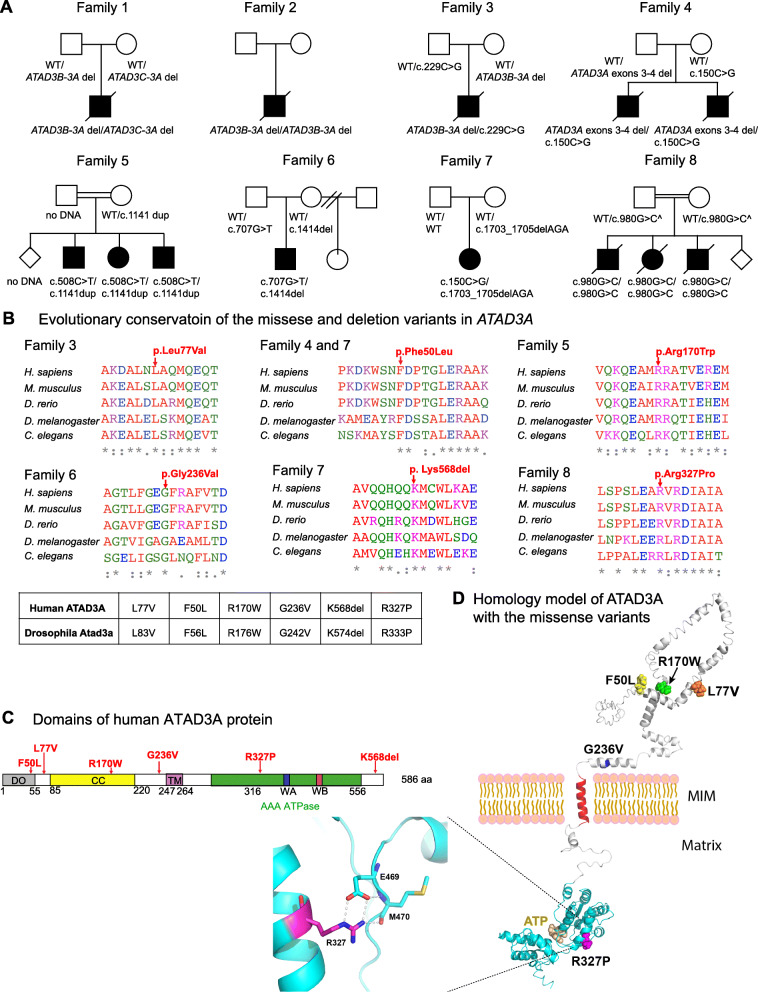
Background: ATPase family AAA-domain containing protein 3A (ATAD3A) is a nuclear-encoded mitochondrial membrane-anchored protein involved in diverse processes including mitochondrial dynamics, mitochondrial DNA organization, and cholesterol metabolism. Biallelic deletions (null), recessive missense variants (hypomorph), and heterozygous missense variants or duplications (antimorph) in ATAD3A lead to neurological syndromes in humans.

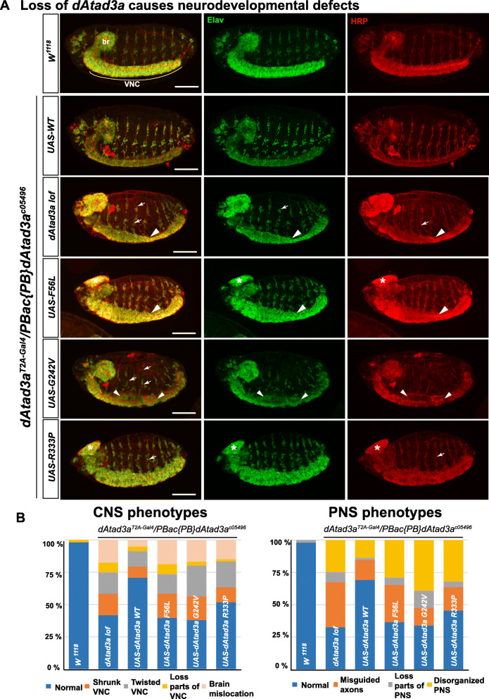
Methods: To expand the mutational spectrum of ATAD3A variants and to provide functional interpretation of missense alleles in trans to deletion alleles, we performed exome sequencing for identification of single nucleotide variants (SNVs) and copy number variants (CNVs) in ATAD3A in individuals with neurological and mitochondrial phenotypes. A Drosophila Atad3a Gal4 knockin-null allele was generated using CRISPR-Cas9 genome editing technology to aid the interpretation of variants.

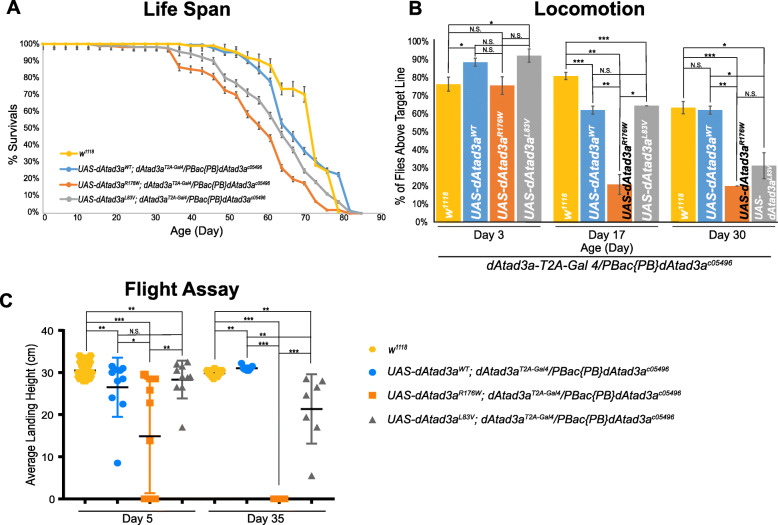
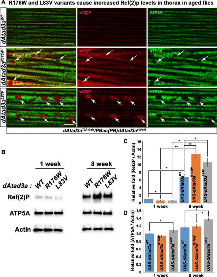
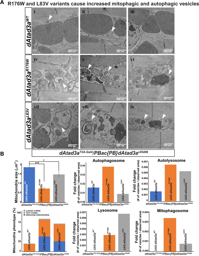
Results: We report 13 individuals from 8 unrelated families with biallelic ATAD3A variants. The variants included four missense variants inherited in trans to loss-of-function alleles (p.(Leu77Val), p.(Phe50Leu), p.(Arg170Trp), p.(Gly236Val)), a homozygous missense variant p.(Arg327Pro), and a heterozygous non-frameshift indel p.(Lys568del). Affected individuals exhibited findings previously associated with ATAD3A pathogenic variation, including developmental delay, hypotonia, congenital cataracts, hypertrophic cardiomyopathy, and cerebellar atrophy. Drosophila studies indicated that Phe50Leu, Gly236Val, Arg327Pro, and Lys568del are severe loss-of-function alleles leading to early developmental lethality. Further, we showed that Phe50Leu, Gly236Val, and Arg327Pro cause neurogenesis defects. On the contrary, Leu77Val and Arg170Trp are partial loss-of-function alleles that cause progressive locomotion defects and whose expression leads to an increase in autophagy and mitophagy in adult muscles.

Conclusion: Our findings expand the allelic spectrum of ATAD3A variants and exemplify the use of a functional assay in Drosophila to aid variant interpretation.

Keywords: AAA+ protein; ATAD3A; Autophagy; Autosomal recessive; Disease; Drosophila; Mitochondria; Neurogenesis.


Conflict of interest statement

J.R.L. has stock ownership in 23andMe, is a paid consultant for Regeneron Pharmaceuticals, and is a co-inventor on multiple US and European patents related to molecular diagnostics for inherited neuropathies, eye diseases, and bacterial genomic fingerprinting. The Department of Molecular and Human Genetics at Baylor College of Medicine receives revenue from clinical genetic testing conducted at Baylor Genetics (BG) Laboratories. J.R.L. serves on the Scientific Advisory Board of BG. KGM is an employee of GeneDx, Inc. The remaining authors declare that they have no competing interests.

Figures

Fig. 1
Fig. 1
Identification of patients with neurological phenotypes with variants in ATAD3A. a Pedigrees of studied families, indicating biallelic variants in ATAD3A identified in 13 individuals from 8 families. Biallelic deletions were identified in families 1 and 2, loss-of-function alleles (intergenic CNV, intragenic CNV, or frameshift SNV) inherited in trans to a missense variant in families 3–7, and a homozygous missense variant in family 8. ^Parents were tested by indirect segregation analysis using SNP arrays. b Protein sequence alignment in multiple species confirms evolutionary conservation of p.L77V, p.F50L, pR170W, p.G236V, p.K568del, and p.R327P, in both humans and Drosophila. A box shows human SNVs in ATAD3A and the Drosophila dAtad3a variants homologous to the human variants. c Schematic representation of protein domains of human ATAD3A and positions of the SNVs. CC indicates coiled-coil domain. TM indicates putative transmembrane domain. Green indicates AAA+ domain containing Walker A motif (WA) and Walker B motif (WB). d In silico protein structure prediction of ATAD3A shows the position of mutated residues
Fig. 2
Fig. 2
Drosophila Atad3a models show various strength of ATAD3A missense variants. a A schematic of the generation of dAtad3a-T2A-Gal4 by CRISPR-Cas9 gene editing and the translation of a Gal4 protein by a ribosomal skipping mechanism. The location of the attP-SA-T2A-Gal4-polyA-attP cassette insertion into the dAtad3a genomic locus is indicated by the dotted lines. The T2A-Gal4 cassette consists of a splice acceptor (SA, light gray) followed by a ribosomal skipping T2A peptide sequence (pink), a Gal4 coding sequence (green), and a polyadenylation signal (light blue). Two inverted attP sites (blue) are positioned at the 5′- and 3′-end of the cassette. b Expression of UAS-mCD8::GFP under the control of dAtad3a-T2A-Gal4 is monitored in larvae and adult flies. c Complementation test results of dAtad3a-T2A-Gal4 alleles. +, complement; −, failure to complement. dAtad3a-T2A-Gal4 fails to complement a deficiency (Df (3R)Excel7329) that lacks the dAtad3a locus and PBac {PB}dAtad3ac05496 null allele, which were rescued by the expression of wild-type dAtad3a cDNA. These data indicate that dAtad3a-T2A-Gal4 is a loss-of-function mutant. d Western blots for fly heads expressing wild-type dAtad3a-V5 or dAtad3a-V5 carrying the homologous SNV mutations identified from patients. Five replicates were quantified. Error bars indicate SEM. P values were calculated using Student’s t test. ***P < 0.001. N.S. indicates not statistically significant. e The lethality caused by dAtad3a loss was rescued by the expression of wild-type dAtad3a, and dAtad3a carrying L83V or R176W but not by those carrying F56L, G242V, K574del, or R333P
Fig. 3
Fig. 3
dAtad3a lof, F56L, G242V, and R333P variants lead to severe neurodevelopmental defects. a Confocal micrographs of wild-type (w1118), dAtad3a-null mutant embryos and those expressing dAtad3aWT, dAtad3aF56L, dAtad3aG242V, and dAtad3aR333P. Elav (green)-stained neurons and anti-HRP (red)-stained neuronal membranes. Br indicates brain, and VNC indicates ventral nerve cord. Arrowheads indicate shrunken and twisted VNC. Arrows indicate misguided and loss of neurons in the PNS. Scale bars indicate 100 μm. b Quantification of CNS and PNS phenotypes shown in mutant embryos. The numbers of embryos for these analyses are as follows: CNS—w1118 (n = 54), wt (n = 58), null (n= 96), F56L (n = 53), G242V (n = 71), and R333P (n = 60). PNS—w1118 (n = 51), wt (n = 52), null (86), F56L (n = 52), G242V (n = 59), and R333P (n = 44)
Fig. 4
Fig. 4
L83V and R175W variants cause behavioral defects in adult flies. a dAtad3a-null mutant flies expressing L83V, and R176W were short lived compared to wild-type rescue animals and wild-type control (w1118). b dAtad3a-null mutant flies expressing L83V, and R176W exhibited progressive climbing defects compared to wild-type rescue controls and wild-type control (w1118). c dAtad3a-null mutant flies expressing R176W exhibited defects in flight ability on the 5th day of their life and complete failure of flight on the 35th day. dAtad3a mutant flies expressing L83V exhibited progressive decline of flight ability compared to rescue control and wild-type control (w1118). b, c Three biological replicates (25 flies per group) were quantified. Error bars indicate SEM. P values were calculated using Student’s t test. *P < 0.05, **P < 0.01,***P < 0.001. N.S. indicates not statistically significant
Fig. 5
Fig. 5
L83V and R176W variants cause increased p62 levels in the thorax in aged flies. a Confocal micrographs of the thorax muscle from 8-week-old flies—dAtad3a-null mutants expressing dAtad3aWT, dAtad3aR176W, or dAtad3aL83V. ATP5A (green) labels mitochondria. Ref (2) P is the Drosophila homolog of p62 (red). Arrows indicate Ref (2) P signals with the absence of ATP5A signals. Scale bars indicate 100 μm. b Western blots for the protein levels of Ref (2) P, ATP5A, and Actin from dAtad3a mutant fly thoraxes expressing dAtad3aWT, dAtad3aR176W, or dAtad3aL83V (n = 10 per genotype). c Quantification of Ref (2) P and d ATP5A level. Ref (2) P and ATP5A were normalized by Actin. Three biological replicates were quantified. Error bars indicate SEM. P values were calculated using Student’s t test. *P < 0.05, **P < 0.01
Fig. 6
Fig. 6
L83V and R176W variants cause aberrant mitochondrial morphology, increased autophagic and mitophagic vesicles. a Electron micrographs of the thorax muscles from 8-week-old dAtad3a mutant flies expressing dAtad3aWT, dAtad3aR176W, or dAtad3aL83V. Arrows show autophagosomes (i, ii, and v), autolysosomes (v and viii), lysosomes (vi and ix), and mitophagosomes (iv and vii). Scale bars indicate 600 nm. b Quantification of mitochondria size, mitochondria phenotypes, numbers of autophagosome, autolysosome, lysosome, and mitophagosome. The respective number of the vesicles was normalized by observed area (μm2). Error bars indicate SEM. P values were calculated using Student’s t test. *P < 0.05, ***P < 0.001

References

    1. Gilquin B, Taillebourg E, Cherradi N, Hubstenberger A, Gay O, Merle N, Assard N, Fauvarque MO, Tomohiro S, Kuge O, et al. The AAA+ ATPase ATAD3A controls mitochondrial dynamics at the interface of the inner and outer membranes. Mol Cell Biol. 2010;30(8):1984–1996. doi: 10.1128/MCB.00007-10. - DOI - PMC - PubMed
    1. Baudier J. ATAD3 proteins: brokers of a mitochondria-endoplasmic reticulum connection in mammalian cells. Biol Rev Camb Philos Soc. 2018;93(2):827–844. doi: 10.1111/brv.12373. - DOI - PubMed
    1. Goller T, Seibold UK, Kremmer E, Voos W, Kolanus W. Atad3 function is essential for early post-implantation development in the mouse. Plos One. 2013;8(1):e54799. doi: 10.1371/journal.pone.0054799. - DOI - PMC - PubMed
    1. Hoffmann M, Bellance N, Rossignol R, Koopman WJ, Willems PH, Mayatepek E, Bossinger O, Distelmaier F. C. elegans ATAD-3 is essential for mitochondrial activity and development. PLoS One. 2009;4:e7644. doi: 10.1371/journal.pone.0007644. - DOI - PMC - PubMed
    1. Fang HY, Chang CL, Hsu SH, Huang CY, Chiang SF, Chiou SH, Huang CH, Hsiao YT, Lin TY, Chiang IP, Hsu WH, Sugano S, Chen CY, Lin CY, Ko WJ, Chow KC. ATPase family AAA domain-containing 3A is a novel anti-apoptotic factor in lung adenocarcinoma cells. J Cell Sci. 2010;123(7):1171–1180. doi: 10.1242/jcs.062034. - DOI - PubMed

Publication types

MeSH terms

Substances

LinkOut - more resources