. 2022 Sep;106(9):1288-1294.
doi: 10.1136/bjophthalmol-2020-318672. Epub 2021 Apr 12.

Short-term real-world outcomes following intravitreal brolucizumab for neovascular AMD: SHIFT study

Affiliations

Short-term real-world outcomes following intravitreal brolucizumab for neovascular AMD: SHIFT study

Louisa Maria Bulirsch et al. Br J Ophthalmol. 2022 Sep.

Abstract

Background: Brolucizumab has recently been approved in Europe as a novel treatment for patients with neovascular age-related macular degeneration (nAMD). We report on early experiences with real-world outcomes of switch to brolucizumab therapy in previously anti-vascular endothelial growth factor (anti-VEGF)-treated patients.

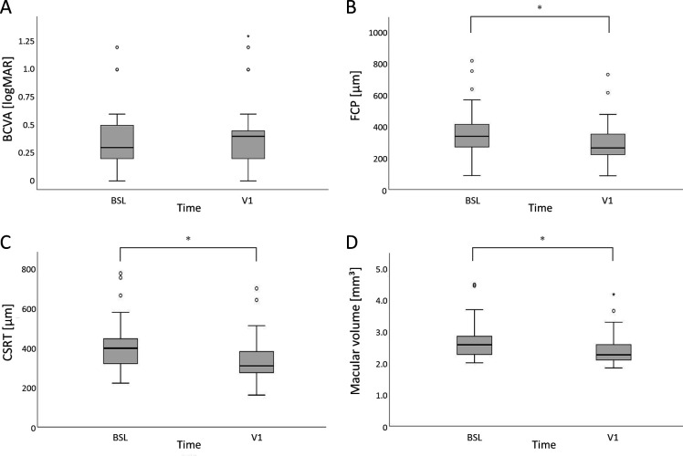
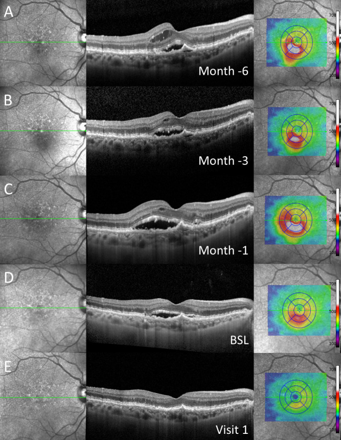
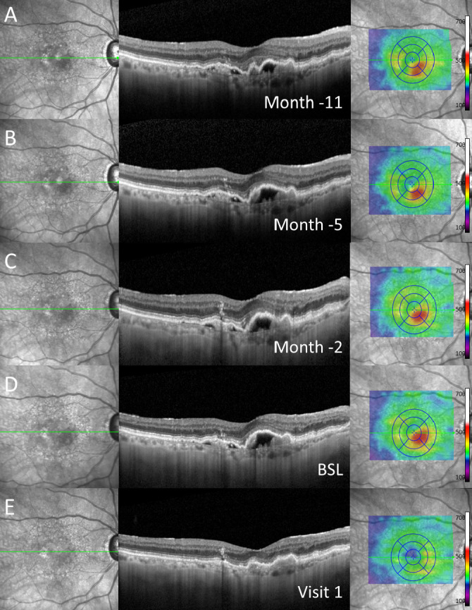
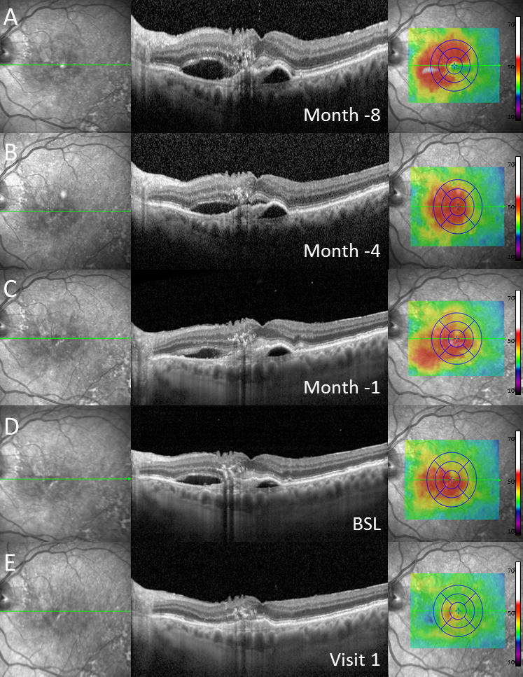
Methods: Patients with recalcitrant nAMD were switched to brolucizumab therapy. Functional and structural parameters 4 weeks after first brolucizumab injection were evaluated including best-corrected visual acuity (BCVA (logMAR)), foveal centre point (FCP (µm)), central subfield retinal thickness (CSRT (µm)) and macular volume (mm³).

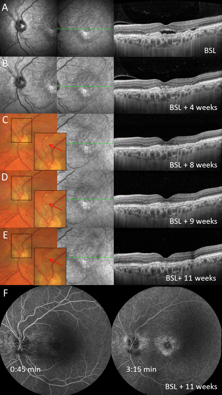
Results: Sixty-three eyes of 57 patients with nAMD (52.6% females) with a mean (±SD) age of 79.5±6.7 years were included. Mean change of BCVA was 0.03±0.14 logMAR (p=0.115). Significant reductions were recorded for FCP with a mean (±SD) change of -66.81±72.63 µm, -66.76±60.71 µm for CSRT and -0.27±0.24 mm³ for macular volume (all p<0.001). Intraocular inflammation was observed in seven eyes of seven patients, including one case of retinal vasculitis.

Conclusions: The results of the SHIFT study indicate that switch to brolucizumab may represent a treatment option in patients with nAMD poorly responsive to other anti-VEGF agents. Further long-term analyses appear prudent to assess efficacy and safety of brolucizumab in a routine clinical setting.

Keywords: Brolucizumab; drugs; imaging; macula; neovascularisation.


Conflict of interest statement

Competing interests: Non-financial support from Heidelberg Engineering to ST, LB, RL, MS and FGH; from CenterVue to ST and FGH; from Optos to ST, LB, RL, MS and FGH; from Carl Zeiss Meditec to ST, LB, RF, FGH and MS. Grant and personal fees from Allergan, Novartis, Bayer and Heidelberg Engineering to ST and FGH; from Apellis, Carl Zeiss Meditec, Acucela, Genentech/Roche, Boehringer-Ingelheim, LIN Bioscience, Pixium, Kanghong, Oxurion, Grayburg Vision, Stealth BioTherapeutics, Geuder and Iveric Bio to FGH. JN: None declared.

Figures

Figure 1
Figure 1
Boxplots (each box: median; 75% and 25% quartiles; whiskers: 10% and 90% quantiles; outliers are defined as sample outliers 1.5 times (circles) and 3 times (star) the IQR above the upper quartile and below the lower quartile) at baseline (BSL) and visit 1 (V1) for (A) best-corrected visual acuity (BCVA (logMAR)), (B) focal centre point (FCP (µm)), (C) central subfield retinal thickness (CSRT (µm)) and (D) macular volume (mm³). Structural outcomes (B–D) showed a significant change after brolucizumab treatment.
Figure 2
Figure 2
Exemplary case of a patient with subretinal fluid at baseline (BSL) (D) and both intraretinal and subretinal fluid 1 (C), 3 (B) and 6 (A) months in historical imaging before baseline despite regular anti-vascular endothelial growth factor (anti-VEGF) treatment as demonstrated in (from left to right) near-infrared imaging, spectral-domain optical coherence tomography through the fovea and colour-coded two-dimensional thickness map for total retinal thickness. One month after switch to brolucizumab (E, visit 1), complete resolution of subretinal fluid was detected. Note: Before switch to brolucizumab, the patient received repetitive, high-frequency intravitreal injections of other anti-VEGF agents over a longer period of time.
Figure 3
Figure 3
Exemplary case of a patient with subretinal pigment epithelial (sub-RPE) fluid at baseline (BSL) (D) and in historical imaging up to 11 months (A–C) before switch to brolucizumab as demonstrated in (from left to right) near-infrared imaging, spectral-domain optical coherence tomography through the fovea and colour-coded two-dimensional thickness map for total retinal thickness. At visit 1 (E), complete resolution of sub-RPE fluid was detected. Note: Before switch to brolucizumab, the patient received repetitive, high-frequency intravitreal injections of other anti-vascular endothelial growth factor agents over a longer period of time.
Figure 4
Figure 4
Exemplary case of a patient with subretinal pigment epithelial (sub-RPE) and subretinal fluid at baseline (BSL) (D) as well as in historical imaging up to 8 months (A–C) before switch to brolucizumab as demonstrated in (from left to right) near-infrared imaging, spectral-domain optical coherence tomography through the fovea and colour-coded two-dimensional thickness map for total retinal thickness. Retinal imaging at visit 1 (E) revealed complete resolution of subretinal and sub-RPE fluid. Note: Before switch to brolucizumab, the patient received repetitive, high-frequency intravitreal injections of other anti-vascular endothelial growth factor agents over a longer period of time.
Figure 5
Figure 5
Exemplary case of a patient with intraretinal and subretinal fluid at baseline (BSL) (D) and in historical images 1 (C), 2 (B) and 6 (A) months before switch to brolucizumab as demonstrated in (from left to right) near-infrared imaging, spectral-domain optical coherence tomography through the fovea and colour-coded two-dimensional thickness map for total retinal thickness. One month after switch (E, visit 1), complete resolution of subretinal and incomplete resolution of intraretinal fluid was demonstrated. Note: Before switch to brolucizumab, the patient received repetitive, high-frequency intravitreal injections of other anti-vascular endothelial growth factor agents over a longer period of time.
Figure 6
Figure 6
Patient with first brolucizumab injection at baseline (BSL, row A), first follow-up visit (visit 1) and second brolucizumab injection 4 weeks later (row B) and development of non-occlusive retinal vasculitis with segmental perivascular sheathing 4 weeks following the second brolucizumab injection (row C). Further follow-up visits (row D–F) as presented by multimodal imaging (from left to right: near-infrared imaging (row A–C) or digital colour fundus photography (row D and E) of the optic nerve head, near-infrared imaging of the macula and spectral-domain optical coherence tomography (SD-OCT) through the fovea). The intra-arterial plaque secondary to retinal vasculitis and its disappearance after treatment initiation is marked by a red arrow in row C, D and E. Seven weeks after first diagnosis of retinal vasculitis, fluorescein angiography did not show signs of vascular occlusion or retinal ischaemia (row F). Of note, subretinal fluid completely resolved after two intravitreal injections of brolucizumab (BSL and V1) in SD-OCT imaging.

References

    1. Lim LS, Mitchell P, Seddon JM, et al. . Age-related macular degeneration. Lancet 2012;379:1728–38. 10.1016/S0140-6736(12)60282-7 - DOI - PubMed
    1. Rosenfeld PJ, Brown DM, Heier JS, et al. . Ranibizumab for neovascular age-related macular degeneration. N Engl J Med 2006;355:1419–31. 10.1056/NEJMoa054481 - DOI - PubMed
    1. Bloch SB, Larsen M, Munch IC. Incidence of legal blindness from age-related macular degeneration in Denmark: year 2000 to 2010. Am J Ophthalmol 2012;153:209–13. 10.1016/j.ajo.2011.10.016 - DOI - PubMed
    1. Holz FG, Tadayoni R, Beatty S, et al. . Multi-country real-life experience of anti-vascular endothelial growth factor therapy for wet age-related macular degeneration. Br J Ophthalmol 2015;99:220–6. 10.1136/bjophthalmol-2014-305327 - DOI - PMC - PubMed
    1. Eghøj MS, Sørensen TL. Tachyphylaxis during treatment of exudative age-related macular degeneration with ranibizumab. Br J Ophthalmol 2012;96:21–3. 10.1136/bjo.2011.203893 - DOI - PubMed

Substances