

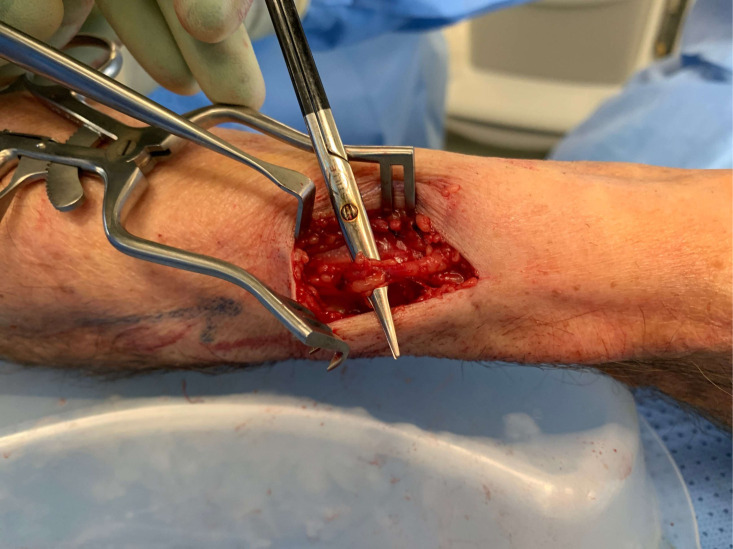
Extradigital glomangiomyoma of the forearm mimicking peripheral nerve sheath tumour and thrombosed varicose vein
- PMID: 33846191
- PMCID: PMC8048004
- DOI: 10.1136/bcr-2020-241221
Extradigital glomangiomyoma of the forearm mimicking peripheral nerve sheath tumour and thrombosed varicose vein
Abstract
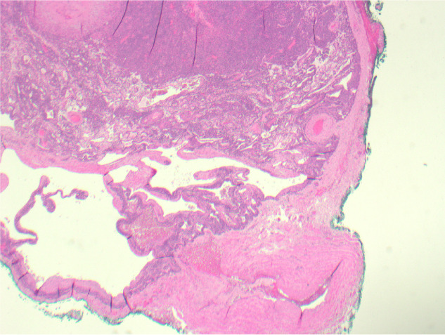
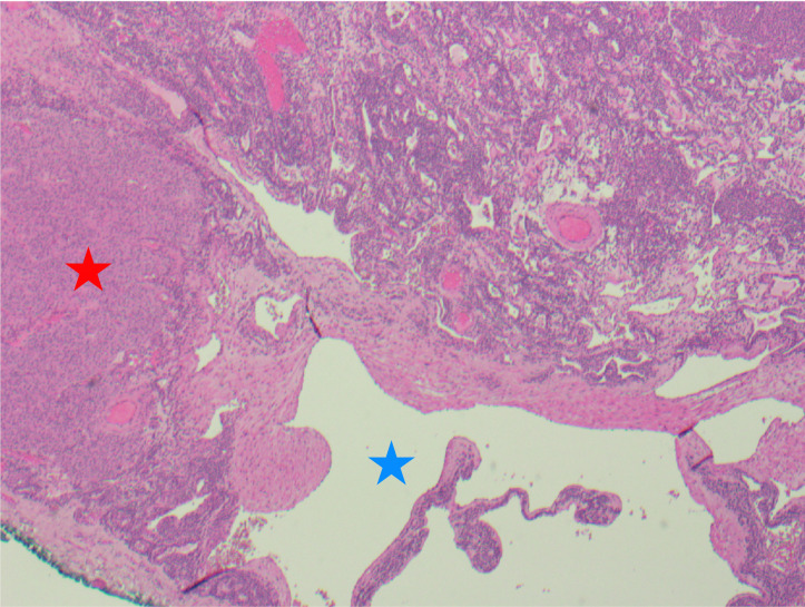
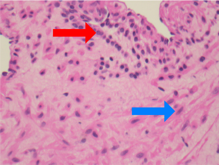
Extradigital glomus tumour is uncommon, little-known outside of its subungual location, and may present without its classic triad of tenderness, cold sensitivity and paroxysmal pain. Imaging is non-specific and diagnosis is often delayed, sometimes for years, leading to unnecessary morbidity. Surgical excision is the treatment of choice, although technique depends on case specifics. Histological subtypes depend on the relative prominence of glomus cells, vascular structures and smooth muscle. The vast majority of glomus tumours are benign. We highlight the importance of considering extradigital glomus tumours when generating differential diagnoses of an atypical painful lesion in a variety of clinical specialties.
Keywords: musculoskeletal and joint disorders; orthopaedic and trauma surgery; plastic and reconstructive surgery.
© BMJ Publishing Group Limited 2021. No commercial re-use. See rights and permissions. Published by BMJ.
Conflict of interest statement
Competing interests: None declared.
Figures
Similar articles
-
Glomus tumour in the forearm: a case report and review of MRI findings.JBR-BTR. 2010 Nov-Dec;93(6):292-5. doi: 10.5334/jbr-btr.342. JBR-BTR. 2010. PMID: 21381525 Review.
-
[Solitary subungual glomangioma].Ann Dermatol Venereol. 2014 Oct;141(10):607-10. doi: 10.1016/j.annder.2014.06.017. Epub 2014 Aug 5. Ann Dermatol Venereol. 2014. PMID: 25288065 French.
-
Acquired Solitary Glomangiomyoma on the Forearm: A Rare Case Report.J Clin Diagn Res. 2016 Jul;10(7):ED10-1. doi: 10.7860/JCDR/2016/19062.8195. Epub 2016 Jul 1. J Clin Diagn Res. 2016. PMID: 27630858 Free PMC article.
-
An Extradigital Glomus Tumor of the Median Antebrachial Vein.J Hand Surg Am. 2018 Jan;43(1):88.e1-88.e4. doi: 10.1016/j.jhsa.2017.07.025. Epub 2017 Sep 6. J Hand Surg Am. 2018. PMID: 28888573
-
Large plaque-like glomangioma in a patient with multiple glomus tumours: review of imaging and histology.Clin Exp Dermatol. 2013 Oct;38(7):693-700. doi: 10.1111/ced.12122. Clin Exp Dermatol. 2013. PMID: 24073652 Review.
Cited by
-
Rapidly progressive malignant glomus tumor of the breast: a case report and review of the literature.J Int Med Res. 2024 Sep;52(9):3000605241272609. doi: 10.1177/03000605241272609. J Int Med Res. 2024. PMID: 39246065 Free PMC article. Review.
-
Glomus Tumor of the Lower Extremity Previously Misdiagnosed as Complex Regional Pain Syndrome in Close Proximity to a Myxofibrosarcoma: A Case Report.J Am Acad Orthop Surg Glob Res Rev. 2022 Jul 6;6(7):e21.00311. doi: 10.5435/JAAOSGlobal-D-21-00311. eCollection 2022 Jul 1. J Am Acad Orthop Surg Glob Res Rev. 2022. PMID: 35797605 Free PMC article.
References
-
- Fletcher CDM, Krishnan Unni K, Mertens F. WHO classification of tumors of soft tissue and bone. ARC Press Lyon, 2002.
Publication types
MeSH terms
LinkOut - more resources
Full Text Sources
Other Literature Sources
Medical