Case Reports
. 2021 Apr 12;14(4):e241591.
doi: 10.1136/bcr-2021-241591.

Dual retrotympanic aural mass

Affiliations
Case Reports

Dual retrotympanic aural mass

Nurul Asma Che Ab Rahim et al. BMJ Case Rep. .

Abstract

High-riding jugular bulb (HRJB), although rare, may pose a challenge as it may be mistaken for other non-alarming condition, such as middle ear effusion. Patients with HRJB classically present with pulsatile tinnitus. We report a unique case of a 26-year-old patient with underlying beta thalassaemia who presented with a 2-month history of intermittent epistaxis and rhinorrhoea. Otoscopic examinations revealed a pulsatile bluish mass behind the right tympanic membrane and a dull left tympanic membrane. Imaging performed revealed a finding of dual retrotympanic pathology, which consisted of a right dehiscent HRJB and left cholesterol granuloma. We highlight a rare case of dual retrotympanic mass as well as its management.

Keywords: ear; nose and throat/otolaryngology; radiology.


Conflict of interest statement

Competing interests: None declared.

Figures

Figure 1
Figure 1
Right otoscopic image showing red-bluish retrotympanic mass occupying the posterior inferior quadrant of the tympanic membrane with some tympanosclerosis.
Figure 2
Figure 2
Otoscopic image of the left ear showing a dull tympanic membrane.
Figure 3
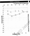
Figure 3
Pure tone audiogram of the right ear showed 10–20 dB conductive hearing loss.
Figure 4
Figure 4
High-resolution CT denotes high, dehiscent right jugular bulb projecting into the right middle ear cavity. Black arrowhead signify Right jugular bulb.
Figure 5
Figure 5
MRI denotes hyperintensity in the T1-weighted soft tissue lesion in the left mastoid and hypotympanum. Black arrowhead signify Left cholesterol granuloma.

Similar articles

References

    1. Sasindran V, Abraham S, Hiremath S, et al. . High-riding jugular bulb: a rare entity. Indian J Otol 2014;20:129–31. 10.4103/0971-7749.136863 - DOI
    1. Moore PJ. The high jugular bulb in ear surgery: three case reports and a review of the literature. J Laryngol Otol 1994;108:772–5. 10.1017/S0022215100128087 - DOI - PubMed
    1. Atilla S, Akpek S, Uslu S, et al. . Computed tomographic evaluation of surgically significant vascular variations related with the temporal bone. Eur J Radiol 1995;20:52–6. 10.1016/0720-048X(95)00619-2 - DOI - PubMed
    1. Ball M, Elloy M, Vaidhyanath R, et al. . Beware the silent presentation of a high and dehiscent jugular bulb in the external ear canal. J Laryngol Otol 2010;124:790–2. 10.1017/S0022215109992349 - DOI - PubMed
    1. Manjila S, Bazil T, Kay M, et al. . Jugular bulb and skull base pathologies: proposal for a novel classification system for jugular bulb positions and microsurgical implications. Neurosurg Focus 2018;45:E5. 10.3171/2018.5.FOCUS18106 - DOI - PubMed

Publication types