. 2021 Apr 12;12(4):387.
doi: 10.1038/s41419-021-03679-8.

ATF3 deficiency impairs the proliferative-secretory phase transition and decidualization in RIF patients

Affiliations

ATF3 deficiency impairs the proliferative-secretory phase transition and decidualization in RIF patients

Zhilong Wang et al. Cell Death Dis. .

Abstract

Decidualization is a complex process involving cellular proliferation and differentiation of the endometrial stroma and is required to establish and support pregnancy. Dysregulated decidualization has been reported to be a critical cause of recurrent implantation failure (RIF). In this study, we found that Activating transcription factor 3 (ATF3) expression was significantly downregulated in the endometrium of RIF patients. Knockdown of ATF3 in human endometrium stromal cells (hESCs) hampers decidualization, while overexpression could trigger the expression of decidual marker genes, and ameliorate the decidualization of hESCs from RIF patients. Mechanistically, ATF3 promotes decidualization by upregulating FOXO1 via suppressing miR-135b expression. In addition, the endometrium of RIF patients was hyperproliferative, while overexpression of ATF3 inhibited the proliferation of hESCs through CDKN1A. These data demonstrate the critical roles of endometrial ATF3 in regulating decidualization and proliferation, and dysregulation of ATF3 in the endometrium may be a novel cause of RIF and therefore represent a potential therapeutic target for RIF.


Conflict of interest statement

The authors declare no competing interests.

Figures

Fig. 1
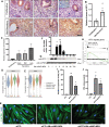
Fig. 1. Aberrant expression of ATF3 in the endometrium of RIF patients.
A, B RNA-seq results showed that ATF3 in the endometrium of RIF patients was downregulated (RIF) compared with that in fertile controls (FER). C ATF3 expression in RIF and FER endometrium was measured by quantitative PCR (qPCR). The expression of ATF3 was normalized to 18S rRNA expression, and the results are presented relative to FER stage (n = 14 for FER group and n = 16 for RIF group). ****P < 0.0001 compared with the FER group. D, E Immunoblotting for ATF3 in the endometrium of RIF patients and the FER group. Quantitative densitometry analysis indicated that ATF3 levels were downregulated in RIF patients (n = 51). ****P < 0.0001 compared with the FER group. F, G Representative immunohistochemical images depicting the expression of ATF3 in the endometrium from fertile women and RIF patients. The negative control (NC) was nonspecific rabbit serum. The H-score of ATF3 expression in the endometrial stromal cells was calculated with IHC-Profiler and ImageJ (scale bar = 50 µm, n = 6, ***P < 0.001). H qPCR was performed to detect the expression of PRL in the endometrium of RIF patients, normalized to 18S rRNA expression (n = 6).
Fig. 2
Fig. 2. The temporal expression and function of ATF3 in the endometrium.
A, B Immunohistochemistry analysis with an anti-ATF3 antibody. Proliferative and mid-secretory phase endometrial tissue samples from normal fertile women. The negative control (NC) was nonspecific rabbit serum. The H-score of ATF3 expression in the endometrial stromal cells was calculated with IHC-Profiler and ImageJ (Scale bar = 50 µm, n = 3, *P < 0.05). C Expression of ATF3 in proliferative and early-, mid-, and late-luteal phase endometrium. Each bar represents an individual biopsy. The data were retrieved from microarray data deposited in the Gene Expression Omnibus (GDS2052). D, E ATF3 expression pattern analyzed with a recent single-cell RNA-seq analysis in stromal cells (D) and all cells (E). The data were retrieved from microarray data deposited in the Gene Expression Omnibus (GSE111976). F The expression pattern of ATF3 in hESCs treated with 0.5 mM 8-Br-cAMP and 1 μM MPA (M + A) for different periods of time (0, 0.5, 1, 2, 4, 8, or 16 h) was evaluated by qPCR. *P < 0.05, **P < 0.01. G The expression pattern of ATF3 in hESCs treated with 0.5 mM 8-Br-cAMP and 1 μM MPA (M + A) for different periods of time (0, 1, 3, 6, 12, 24, 48, or 72 h) was evaluated by western blot. H GSEA results showed that the ATF3-regulated gene term was enriched during in vitro decidualization. I, J hESCs were transfected with siATF3 or siCtl for 48 h and then treated with a decidualization stimulus. The expression and secretion of PRL were measured by qPCR and ELISA, respectively. *P < 0.05, **P < 0.01 compared with the CTL/M + A group (8-Br-cAMP+MPA). K Immunofluorescence was performed to analyze the morphological transformation of hESCs.
Fig. 3
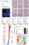
Fig. 3. DEGs in hESCs after knockdown of ATF3 during in vitro decidualization.
A Heat map showing hierarchical clustering of gene expression in decidualization and knockdown of the ATF3 group. B, C KEGG pathway enrichment analysis and GO analysis of the DEGs. D The DEGs related to the altered KEGG pathways. E GSEA results showed that the FoxO-targeted gene term was downregulated when ATF3 was silenced. F The relative expression of FoxO1 target genes, such as PRL, IGFBP-1, DCN, and LEFTY2.
Fig. 4
Fig. 4. ATF3 regulates decidualization mediated by FOXO1.
A hESCs were treated with Ad-LacZ or Ad-ATF3-His for 48 h and transfected with IRS-luc and Rinella for dual-luciferase assays. ****P < 0.0001. BD hESCs were transfected with Ad-ATF3-his (0 or 40 MOI) or Ad-LacZ. si-FOXO1 was used to knockdown the expression of FOXO1, and prolactin secretion was measured by an enzyme-linked fluorescent assay (ELFA). *P < 0.05. Immunofluorescence was performed to analyze the morphological transformation of hESCs. Scale bar = 50 μm. E hESCs were transfected with Ad-ATF3-his (0, 20, or 40 MOI) or Ad-LacZ. FOXO1 and His protein expression levels were measured by western blot.
Fig. 5
Fig. 5. miR-135b mediates the regulation of FOXO1 by ATF3 during decidualization.
A hESCs were transfected with Ad-ATF3-his (0 or 40 MOI) or Ad-LacZ. si-Dicer was used to knockdown the expression of Dicer, and prolactin secretion was measured by ELFA. *P < 0.05, **P < 0.01, ***P < 0.001. B, C miRNA array and qPCR were performed to detect the differentially expressed miRNAs in RIF patients. Data were downloaded from the GEO database (GSE71332). The expression of miR-135b was validated by qPCR and normalized to U6 expression (n = 9 for FER group and 12 for RIF group, *P < 0.05). D The correlation of ATF3 expression and miR-135b with the data in B. E hESCs were transfected with Ad-ATF3-his (0, 20, or 40 MOI) or Ad-LacZ. miR-135b expression levels were analyzed by qPCR. *P < 0.05. F GSEA results showed that the AAGCCAT miR-135B term was activated after overexpression of ATF3. G hESCs were treated with 8-Br-cAMP+MPA for different times, and then the expression of miR-135b was measured by qPCR. ***P < 0.001, ****P < 0.0001. H ChIP-PCR was used to detect the binding site of ATF3 on the promotor of miR-135b. I hESCs were transfected with miR-135b mimic (10 nM) or mimic control. After 24 h, the cells were treated with 0.5 mM 8-Br-cAMP and 1 μM MPA for an additional 3 days. Prolactin released into the medium was measured by ELFA. *P < 0.05, **P < 0.01, ***P < 0.001. J hESCs were transfected with hsa-miR-135b mimic (0, 5, or 10 nM) or mimic control. FOXO1 protein levels were analyzed by western blot. **P < 0.01. K, L Putative 7 bp paired miR-135b/a-target sites in the 3′UTR of human FOXO1 mRNA. Analysis of miR-135b modulation of the wild-type and mutant FOXO1 3′ UTR luciferase reporter activity. *P < 0.05, ns not significant.
Fig. 6
Fig. 6. ATF3 regulates the cell cycle of hESCs.
A GSEA results showed that the cell cycle term was activated in the endometrium of RIF patients (NES: 1.644476, P = 1.023820e−03). B. MKI67 staining was used to measure the proliferation of stromal cells in the endometrium of RIF patients and fertile controls (ST, stromal cells, GE, glandular epithelium, bar = 50 µm, n = 5). C immunofluorescence was performed to determine the expression of ATF3 and MKI67 in endometrium from fertile controls and RIF patients. D The cell cycle-regulated genes were measured by qPCR in the endometrium of RIF patients and fertile controls, n = 6. E TC-seq analysis was performed to analyze the DEGs in the control, decidualization, and siATF3+decidualization groups. Genes in clusters 1 and 6 were upregulated by siATF3, while those in cluster 2 were downregulated. F The correlation of ATF3 expression and CDKN1A. G hESCs were treated with Ad-ATF3-His or Ad-LacZ, and a CCK-8 assay was performed to detect the cell proliferation rate. H Genes related to cell proliferation and decidualization change regularly during in vitro-induced decidualization.
Fig. 7
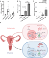
Fig. 7. Decidualization is augmented by overexpression of ATF3 in RIF patients.
A hESCs (from fertile controls and RIF patients) were treated with 8-Br-cAMP+MPA for 3 days. Prolactin secretion was measured by ELFA. n = 5, *P < 0.05, **P < 0.01. B, C hESCs from RIF patients were infected with Ad-ATF3-his (0 or 40 MOI) or Ad-LacZ for 48 h followed by treatment with 0.5 mM 8-Br-cAMP and 1 μM MPA for 3 days. Prolactin released into the medium was measured by ELFA and qPCR. n = 4, *P < 0.05, **P < 0.01. D Schematic representation of the role of ATF3 in the regulation of decidualization in RIF patients and fertile control groups.

Similar articles

Cited by

References

    1. Wang H, Dey SK. Roadmap to embryo implantation: clues from mouse models. Nat. Rev. Genet. 2006;7:185–199. doi: 10.1038/nrg1808. - DOI - PubMed
    1. Matsumoto L, Hirota Y, Saito-Fujita T, Takeda N, Tanaka T, Hiraoka T, et al. HIF2alpha in the uterine stroma permits embryo invasion and luminal epithelium detachment. J. Clin. Invest. 2018;128:3186–3197. doi: 10.1172/JCI98931. - DOI - PMC - PubMed
    1. European IVFmC, European Society of Human R, Embryology. Calhaz-Jorge C, De Geyter C, Kupka MS, et al. Assisted reproductive technology in Europe, 2013: results generated from European registers by ESHRE. Hum. Reprod. 2017;32:1957–1973. doi: 10.1093/humrep/dex264. - DOI - PubMed
    1. Aikawa S, Kano K, Inoue A, Wang J, Saigusa D, Nagamatsu T, et al. Autotaxin-lysophosphatidic acid-LPA3 signaling at the embryo-epithelial boundary controls decidualization pathways. EMBO J. 2017;36:2146–2160. doi: 10.15252/embj.201696290. - DOI - PMC - PubMed
    1. Gu XW, Yang Y, Li T, Chen ZC, Fu T, Pan JM, et al. ATP mediates the interaction between human blastocyst and endometrium. Cell Prolif. 2020;53:e12737. - PMC - PubMed

Publication types

MeSH terms