. 2021 Apr 12;4(1):475.
doi: 10.1038/s42003-021-02030-3.

Human ACE2 receptor polymorphisms and altered susceptibility to SARS-CoV-2

Affiliations

Human ACE2 receptor polymorphisms and altered susceptibility to SARS-CoV-2

Kushal Suryamohan et al. Commun Biol. .

Abstract

COVID-19 is a respiratory illness caused by a novel coronavirus called SARS-CoV-2. The viral spike (S) protein engages the human angiotensin-converting enzyme 2 (ACE2) receptor to invade host cells with ~10-15-fold higher affinity compared to SARS-CoV S-protein, making it highly infectious. Here, we assessed if ACE2 polymorphisms can alter host susceptibility to SARS-CoV-2 by affecting this interaction. We analyzed over 290,000 samples representing >400 population groups from public genomic datasets and identified multiple ACE2 protein-altering variants. Using reported structural data, we identified natural ACE2 variants that could potentially affect virus-host interaction and thereby alter host susceptibility. These include variants S19P, I21V, E23K, K26R, T27A, N64K, T92I, Q102P and H378R that were predicted to increase susceptibility, while variants K31R, N33I, H34R, E35K, E37K, D38V, Y50F, N51S, M62V, K68E, F72V, Y83H, G326E, G352V, D355N, Q388L and D509Y were predicted to be protective variants that show decreased binding to S-protein. Using biochemical assays, we confirmed that K31R and E37K had decreased affinity, and K26R and T92I variants showed increased affinity for S-protein when compared to wildtype ACE2. Consistent with this, soluble ACE2 K26R and T92I were more effective in blocking entry of S-protein pseudotyped virus suggesting that ACE2 variants can modulate susceptibility to SARS-CoV-2.


Conflict of interest statement

Employees of ModMab Therapeutics and MedGenome hold shares/options in their respective companies. The remaining authors declare no competing interests.

Figures
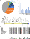
Fig. 1
Fig. 1. ACE2 polymorphisms.
a Pie chart representing protein altering variations in ACE2 by allele count and source. b Log base 10 pseudo count adjusted (+1) observed ACE2 allele counts of mutants predicted to impact S-protein binding. Singletons are marked with a ^ and direct S-protein contact residues are underlined. c ACE2 protein domain showing positions with polymorphisms that can alter SARS-CoV-2 S-protein binding. Recurrent polymorphisms (n > 1) that were predicted to not impact S-protein binding are shown in light grey. Residues within the ACE2 peptidase domain (PD) known to interact with viral S-protein are shown as red vertical lines within the peptidase domain in the ACE2 diagram. d Multiple sequence alignment of the S-protein interacting ACE2 sequence from indicated species. ACE2 NxT/S glycosylation motif disrupted in dog, rat, palm civet, and several bat ACE2 is highlighted in red. ACE2 residues that mediate contact with NL63-CoV, SARS-CoV and SARS-CoV-2 are shown as blue, green and orange bars, respectively.
Fig. 2
Fig. 2. Human ACE2 polymorphisms mapped to the structure of human ACE2 in complex with the SARS-CoV-2 RBD.
Residues in ACE2 showing polymorphic variation in human populations were mapped on to the structure of the ACE2/SARS-CoV-2 RBD (PDB: 6VW1) and colored according to their effect on the predicted affinity to SARS-CoV-2 RBD. Polymorphisms that were predicted to enhance the binding between ACE2 and the S-protein are colored in magenta. Polymorphisms that are predicted to disrupt the binding between ACE2 and the S-protein are colored in dark blue. The variable loop in the ridge binding motif consisting of residues V483 and E484 is shown in red. This region in the structure (PDB: 6LZG) is zoomed-in to show variants predicted to enhance or disrupt the ACE2 – SARS-CoV-2 interaction.
Fig. 3
Fig. 3. Structural basis of interaction between human ACE2 polymorphic variants and SARS-CoV-2 S-protein.
a K26R, b T92I, c K31R and d E37K. Interaction mapping done using PDB structure 6VW1.
Fig. 4
Fig. 4. Binding affinity of SARS-CoV-2 S-RBD, S1 and S-trimer.
ac ELISA assay measuring the affinity of indicated ACE2 WT or variants for SARS-CoV-2 S-RBD (a), S1 (b) and S-trimer (c). di Sensorgrams for the binding of a 3-fold dilution series of monomeric and dimeric ACE2 variants to streptavidin-captured SARS-CoV-2 RBD are provided (black). Fits to the empirical binding curves are shown overlaid (red).
Fig. 5
Fig. 5. ACE2 variants block pseudotyped SARS-CoV-2 virus infection.
Inhibition of pseudovirus entry into HEK293T cells by ACE2 WT and mutants S19P, T92I, and K26R. A range of seven different concentrations of Fc-tagged ACE2 proteins were mixed with pseudoviruses and the infectivity was measured by the luciferase signal as an indication of the amount of virus entering the cells. Data are presented as the mean ± SD (standard deviation). Sample size n = 3.

References

    1. Li F. Structure, function, and evolution of coronavirus spike proteins. Annu. Rev. Virol. 2016;3:237–261. doi: 10.1146/annurev-virology-110615-042301. - DOI - PMC - PubMed
    1. Graham RL, Baric RS. Recombination, reservoirs, and the modular spike: mechanisms of coronavirus cross-species transmission. J. Virol. 2010;84:3134–3146. doi: 10.1128/JVI.01394-09. - DOI - PMC - PubMed
    1. Holmes KV. SARS-associated coronavirus. N. Engl. J. Med. 2003;348:1948–1951. doi: 10.1056/NEJMp030078. - DOI - PubMed
    1. Zaki AM, van Boheemen S, Bestebroer TM, Osterhaus AD, Fouchier RA. Isolation of a novel coronavirus from a man with pneumonia in Saudi Arabia. N. Engl. J. Med. 2012;367:1814–1820. doi: 10.1056/NEJMoa1211721. - DOI - PubMed
    1. Zhu N, et al. A novel coronavirus from patients with pneumonia in China, 2019. N. Engl. J. Med. 2020;382:727–733. doi: 10.1056/NEJMoa2001017. - DOI - PMC - PubMed

MeSH terms

Substances