. 2021 Jul;14(4):949-962.
doi: 10.1038/s41385-021-00402-w. Epub 2021 Apr 12.

Control of IFN-I responses by the aminopeptidase IRAP in neonatal C57BL/6 alveolar macrophages during RSV infection

Affiliations

Control of IFN-I responses by the aminopeptidase IRAP in neonatal C57BL/6 alveolar macrophages during RSV infection

Carole Drajac et al. Mucosal Immunol. 2021 Jul.

Abstract

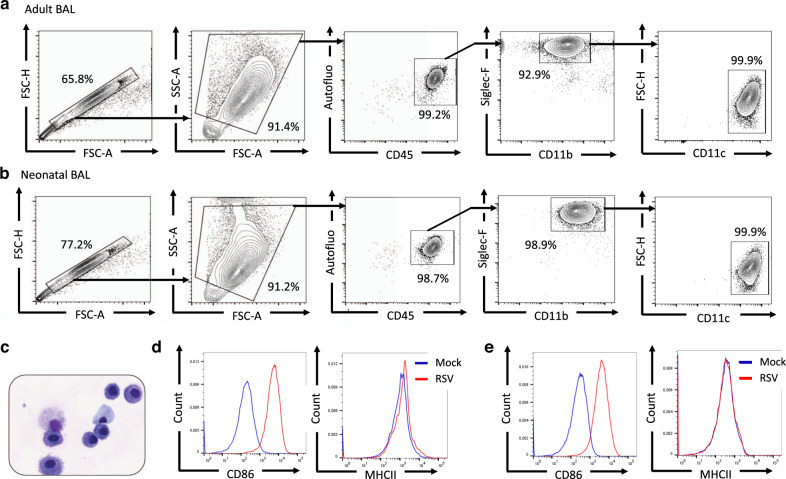
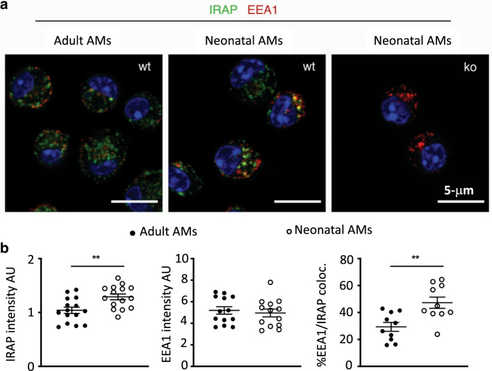
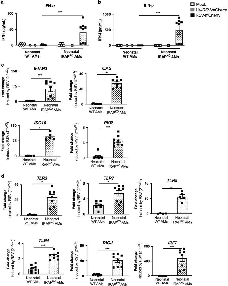
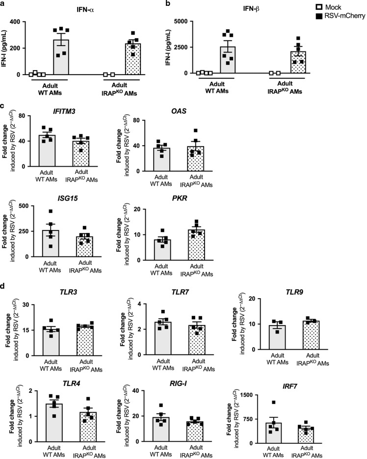
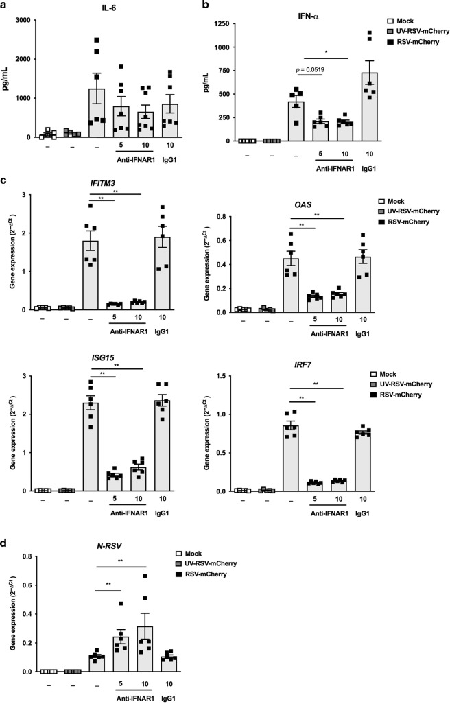
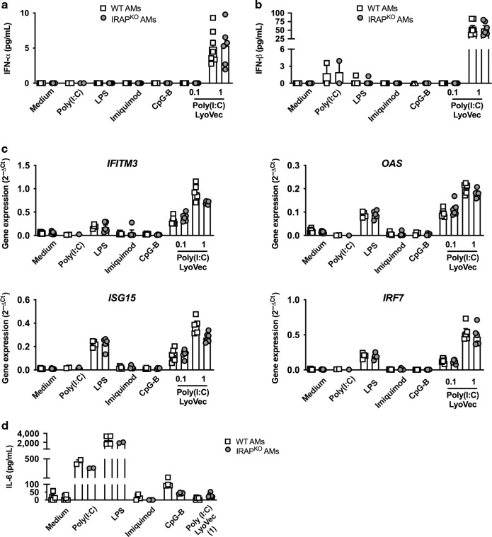
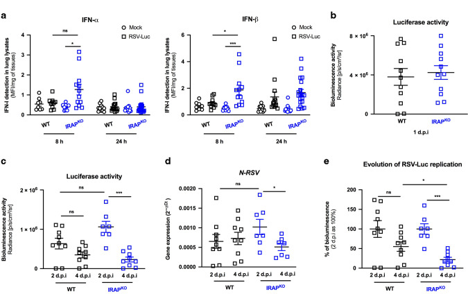
Respiratory Syncytial Virus (RSV) is the major cause of lower respiratory tract infection in infants, in whom, the sensing of RSV by innate immune receptors and its regulation are still poorly described. However, the severe bronchiolitis following RSV infection in neonates has been associated with a defect in type I interferons (IFN-I) production, a cytokine produced mainly by alveolar macrophages (AMs) upon RSV infection in adults. In the present study, neonatal C57BL/6 AMs mobilized very weakly the IFN-I pathway upon RSV infection in vitro and failed to restrain virus replication. However, IFN-I productions by neonatal AMs were substantially increased by the deletion of Insulin-Responsive AminoPeptidase (IRAP), a protein previously involved in the regulation of IFN-I production by dendritic cells. Moreover, neonatal IRAPKO AMs showed a higher expression of IFN-stimulated genes than their wild-type C57BL/6 counterpart. Interestingly, depletion of IRAP did not affect adult AM responses. Finally, we demonstrated that newborn IRAPKO mice infected with RSV had more IFN-I in their lungs and eliminated the virus more efficiently than WT neonates. Taken together, early-life susceptibility to RSV infection may be related to an original age-dependent suppressive function of IRAP on the IFN-I driven-antiviral responses in neonatal AMs.


Conflict of interest statement

The author declares no competing interests.

Figures

Fig. 1
Fig. 1. Neonatal C57BL/6 mice are permissive to RSV infection and are unable to active an efficient type I IFN pathway.
Six days old neonates and 8 weeks old adult C57BL/6 mice were infected i.n. with rRSV-Luc (1.75 × 106 pfu/mL, 10 µL/neonate, 50 µL/adult). a Quantification of luciferase activity in lung and NT homogenates (radiance in photons/s/cm2/sr/mg of tissues) at 2 and 4 days post-infection (d.p.i.). Results are expressed as mean of individual ± SEM (one representative experiment of two with n = 4–6 mice/group, *P < 0.05; **P < 0.01). b IFN-β production was measured by luminex assays in lung homogenates at 1 d.p.i. c The expressions of different ISGs and innate sensing receptors were determined by qRT-PCR on lung homogenates at 1 d.p.i. The level of expression was calculated by the formula 2−ΔCt with ΔCTRSV = Ctgene − CtHouse keeping-genes. Results are expressed in fold-change determined with the formula 2−ΔΔCt with ΔΔCT = ΔCTRSV/ΔCTmock. Data are from two experiments. Means ± SEM are represented. b, c Results are expressed as mean of individual ± SEM (one representative experiment of three with n = 3–7 mice/group, *P < 0.05; **P < 0.01).
Fig. 2
Fig. 2. Adult or neonatal AMs collected from BAL of neonatal C57BL6 mice are activated upon RSV infection.
Cells were collected from BAL of adult (a, d) or 6 days old (b–e) C57BL/6 mice and analyzed for phenotype by flow cytometry. Representative contour plots of adult AMs (a) or 6 days old mice (b) are shown. c Representative cytospin picture of BAL collected from 6 days old mice and stained with May-Grunwald Giemsa coloration. d, e AMs collected from BAL of adult animals (d) or 6 days old C57BL/6 mice (e) were plated for 24 h then infected for 24 h with RSV (MOI = 5) or Mock control. Expression level (MFI level) of CD86 or MHCII activation markers was analyzed at the cell surface of neonatal AMs by flow cytometry.
Fig. 3
Fig. 3. Intracellular localization of IRAP in adult or neonatal AMs.
AMs collected from BAL of adult C57BL/6 mice or 6 days old C57BL/6 mice or 6 days old IRAP-deficient mice were plated for 24 h. a Cells were fixed and stained for IRAP (Green), early endosome antigen 1 (EEA1), an early endosomal marker (red), and nuclear DAPI labeling (blue). Bars represent 5 μm scale. b The quantification of IRAP and early endosomal marker (EEA1) and their co-localization were performed (n = 15 cells for each labeling). Image treatment and analysis were performed with ImageJ software. Means ± SEM are represented. **P < 0.01.
Fig. 4
Fig. 4. Deletion of IRAP restores type I IFN pathway in neonatal AMs following in vitro RSV infection.
AMs isolated from BAL of neonatal C57BL/6 wild-type (WT) or IRAP-deficient (IRAPKO) mice were exposed to Hep2-supernatant (Mock, white symbol) or rRSV-mCherry (RSV-mCherry, black symbol) or UV-inactivated rRSV-mCherry (UV-RSV-mCherry, gray symbol) at MOI of 5 for 2 h. The productions of IFN-α (a) and IFN-β (b) were measured 24 h post-infection in supernatants using ProcartaxPlex immunoassay. The expressions of different ISGs (c) and innate sensing receptors (d) were determined by qRT-PCR on cell lysates 24 h post-infection. The level of expression was calculated by the formula 2−ΔCt with ΔCTRSV = Ctgene − CtGADPH. Results are expressed in fold-change determined with the formula 2−ΔΔCt with ΔΔCT = ΔCTRSV/ΔCTmock. Data are from two experiments. Means ± SEM are represented. *P < 0.05; **P < 0.01; ***P < 0.001.
Fig. 5
Fig. 5. Deletion of IRAP in adult AMs does not affect the type I IFN response to in vitro RSV infection.
Adult AMs from WT C57BL/6 or IRAP-deficient (IRAPKO) mice were exposed to Hep2-supernatant (Mock, white symbol) or rRSV-mCherry (RSV-mCherry, black symbol) at MOI of 5 for 2 h. The productions of IFN-α (a) and IFN-β (b) were measured 24 h post-infection in supernatants using ProcartaxPlex immunoassay. The expressions of different ISGs (c) and innate sensing receptors (d) were determined by qRT-PCR on cell lysates 24 h post-infection. The level of expression was calculated by the formula 2−ΔCt with ΔCTRSV = Ctgene − CtGADPH. Results are expressed in fold-change determined with the formula 2−ΔΔCt with ΔΔCT = ΔCTRSV/ΔCTmock. Data are from two experiments. Means ± SEM are represented.
Fig. 6
Fig. 6. RSV replication in AMs is retrained by induction of type I IFN signaling.
a–d Adult AMs collected from BAL of WT C57BL/6 mice were exposed to Hep2-supernatant (Mock, white symbol) or rRSV-mCherry (RSV-mCherry, black symbol) or UV-inactivated rRSV-mCherry (UV-RSV-mCherry, gray symbol) at MOI of 5 for 2 h in the presence or not of anti-IFNα/β receptor (IFNAR) antibody (5 or 10 μg/mL). The productions of IL-6 (a) or IFN-α (b) were measured 24 h post-infection in supernatants using ELISA or ProcartaxPlex immunoassay, respectively. c The expressions of different ISGs were determined by qRT-PCR on cell lysates 24 h post-infection. d RSV replication was evaluated by qRT-PCR of N-RSV gene expression on cell lysates 24 h post-infection. The level of expression was calculated by the formula 2−ΔCt with ΔCT = CtRSV − CtGADPH. d Data are from two experiments. Means ± SEM are represented. *P < 0.05; **P < 0.01; ***P < 0.001.
Fig. 7
Fig. 7. Neonatal IRAPKO AMs restrain more efficiently the RSV replication than neonatal WT AMs.
Neonatal or adult AMs collected from BAL of wild-type (WT) C57BL/6 mice or IRAP-deficient (IRAPKO) mice were exposed to rRSV-mCherry (RSV-mCherry, black symbol) at MOI of 5 or Hep2-supernatant (Mock, white symbol) for 2 h. N-RSV RNA expression was quantified by qRT-PCR on cell lysates 24 h post-infection. Results were calculated by the formula 2−ΔCt with ΔCT = CtRSV − CtGADPH. Data are from two experiments. Means ± SEM are represented. **P < 0.01; ***P < 0.001.
Fig. 8
Fig. 8. Type I IFN and inflammatory responses upon specific innate sensing receptor stimulation do not depend upon IRAP expression in neonatal AMs.
Neonatal AMs from wild-type (WT, white square) or IRAP-deficient (IRAPKO, gray circle) mice were stimulated with different PAMPs: Poly(I:C) (100 μg/mL), LPS (10 µg/mL), Imiquimod (100 μg/mL), CpG-B (100 μg/mL), or Poly(I:C)-LyoVec (0.1 or 1 μg/mL). The productions of IFN-α (a) and IFN-β (b) were measured 24 h post-infection in supernatants using ProcartaxPlex immunoassay. c The expression of different ISGs was determined by qRT-PCR performed on cell lysates 24 h post-infection. The level of expression was calculated by the formula 2−ΔCt with ΔCT = Ctgene − CtGADPH. Data are from four experiments. Means ± SEM are represented. d The production of IL-6 was measured 24 h post-stimulation in supernatants using ELISA. Data are from four experiments. Means ± SEM are represented.
Fig. 9
Fig. 9. IRAP-deficient newborn mice display an enhanced production of type I IFNs in the lungs and better control of RSV replication.
Wild-type (WT) or IRAP-deficient (IRAPKO) neonatal mice were infected with Hep2-supernatant (Mock) or rRSV-Luc (RSV-Luc). a The productions of IFN-α and IFN-β were measured post-infection in lung lysates using ProcartaxPlex immunoassay. Results are expressed in mean fluorescent intensity (MFI) per mg of lung lysates at 8 or 24 h post-infection. b, c Luciferase activity associated to viral replication was measured in lung lysates (left lung lobe) by quantification of photon emission (radiance in photon/sec/cm2/sr). d N-RSV gene expression was measured in the lung lysates by qRT-PCR and calculated by the formula 2−ΔCt with ΔCT = CtN-RSV − CtGADPH. e Reduction of RSV replication between 2 d.p.i and 4 d.p.i. Each individual bioluminescence signal obtained at 2 d.p.i and 4 d.p.i was normalized to the mean bioluminescence value of each group at 2 d.p.i considered as 100%. a Data are mean ± SEM from three independent experiments with n = 8–14 mice per group and timepoints. b–e Data are mean ± SEM from two independent experiments with n = 7–11 mice per group and timepoints. *P < 0.05; **P < 0.01; ***P < 0.001.

Similar articles

Cited by

References

    1. Smyth RL, Openshaw PJ. Bronchiolitis. Lancet. 2006;368:312–322. - PubMed
    1. Janssen R, et al. Genetic susceptibility to respiratory syncytial virus bronchiolitis is predominantly associated with innate immune genes. J. Infect. Dis. 2007;196:826–834. - PubMed
    1. Lambert L, Sagfors AM, Openshaw PJ, Culley FJ. Immunity to RSV in early-life. Front Immunol. 2014;5:466. - PMC - PubMed
    1. Cormier SA, You D, Honnegowda S. The use of a neonatal mouse model to study respiratory syncytial virus infections. Expert Rev. Anti Infect. Ther. 2010;8:1371–1380. - PMC - PubMed
    1. de Kleer IM, et al. Perinatal activation of the interleukin-33 pathway promotes Type 2 immunity in the developing lung. Immunity. 2016;45:1285–1298. - PubMed

Publication types

MeSH terms