. 2021 Apr 12;4(1):478.
doi: 10.1038/s42003-021-01944-2.

Deletion of LBR N-terminal domains recapitulates Pelger-Huet anomaly phenotypes in mouse without disrupting X chromosome inactivation

Affiliations

Deletion of LBR N-terminal domains recapitulates Pelger-Huet anomaly phenotypes in mouse without disrupting X chromosome inactivation

Alexander Neil Young et al. Commun Biol. .

Abstract

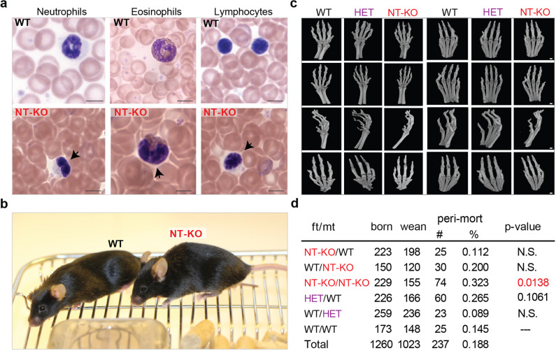
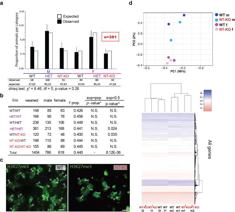
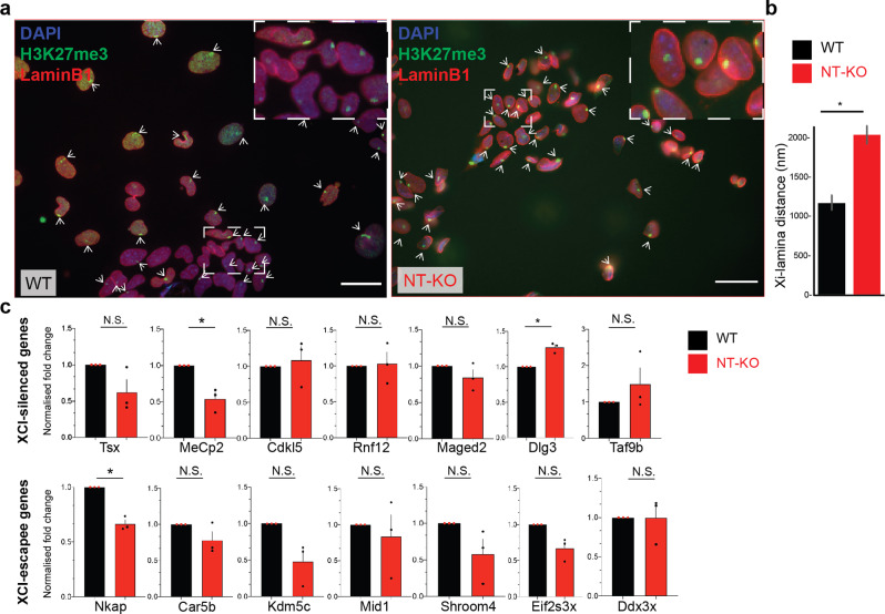
Mutations in the gene encoding Lamin B receptor (LBR), a nuclear-membrane protein with sterol reductase activity, have been linked to rare human disorders. Phenotypes range from a benign blood disorder, such as Pelger-Huet anomaly (PHA), affecting the morphology and chromatin organization of white blood cells, to embryonic lethality as for Greenberg dysplasia (GRBGD). Existing PHA mouse models do not fully recapitulate the human phenotypes, hindering efforts to understand the molecular etiology of this disorder. Here we show, using CRISPR/Cas-9 gene editing technology, that a 236bp N-terminal deletion in the mouse Lbr gene, generating a protein missing the N-terminal domains of LBR, presents a superior model of human PHA. Further, we address recent reports of a link between Lbr and defects in X chromosome inactivation (XCI) and show that our mouse mutant displays minor X chromosome inactivation defects that do not lead to any overt phenotypes in vivo. We suggest that our N-terminal deletion model provides a valuable pre-clinical tool to the research community and will aid in further understanding the etiology of PHA and the diverse functions of LBR.


Conflict of interest statement

The authors declare no competing interests.

Figures

Fig. 1
Fig. 1. LBR NT-KO mice show laminopathy defects and increased perinatal mortality but not skin or skeletal defects.
a Top, Pelguer–Huet characteristic neutrophil and eosinophil and lymphocyte chromatin defects are shown (blood from female animals are presented, but no sex-specific effect has been observed). Black arrows in LBR NT-KO animals indicate defects in chromatin organization in the shown cell types. Scale bar indicates 5 µm. b No skin/fur defects were observed in LBR NT-KO animals. A female adult WT and a LBR NT-KO mouse are shown next to each other are shown. c No skeletal defects in front (left) and rear (right) paws have been observed in any analyzed category (female animals are shown in this figure, but no sex-specific effect has been observed). Scale bar indicates 1 mm. d The table shows perinatal mortality from different classes of crosses indicating the parental origin of the mutation (father: ft; mother: mt). born: born; wean: weaned; peri-mort: perinatal mortality; mutation classes are shown. A one-way ANOVA (Kruskal–Wallis test), followed by a post hoc multiple comparison tests (Dunn’s test) have been used to compare the number of weaned mice in each group to the WT/WT condition (the analysis was done per litter).
Fig. 2
Fig. 2. A N-term LBR deletion, does not affect XCI in vivo.
a 34 Lbr heterozygous (Lbr+/Lbr236, HET) mice pairs were crossed and the offspring have been genotyped by PCR analysis (see schematic in Supplementary Fig. 1a). Observed offspring genotypes indicated in black with error bars indicating 95% confidence interval (CI). A significant skewing against females is predicted in the analyzed offspring, in case Lbr plays a major role in XCI in vivo (M: male; F: female). Chi-square test analysis reveals no significant differences between expected and observed classes of male and female offspring (X2 = 6.46, df = 5, p-value = 0.26). The number of animals used is shown in red (n = 381). b The table shows different classes of crosses and indicates the parental origin and type of the mutation. Sex ratio and binomial test analysis are also shown. Statistical tests are run vs the observed total population ratio of males/females (m/f) and vs the expected, theoretical 50% ratio. *Two-tailed binomial test against population female proportion (0.440). ^Two-tailed binomial test against expected female proportion (0.50). c Xi-lamina association reveals no differences between WT and LBR NT-KO animals. H3K27me3 staining (Xi surrogate) is shown. Cross-sections from thymus tissue are shown. Arrows indicate the inactive X chromosome. Scale bar indicates 10 µm. d Top, RNA-Seq principal component analysis and hierarchical clustering of the LBR NT-KO vs WT are shown (number of genes: 12,791). The heatmap depicts scaled expression for better visualization. Categories are shown in the legends.
Fig. 3
Fig. 3. The reported LBR N-term mutation affects the Xi localization but not gene silencing in random XCI.
a Left, Xi-lamina localization is shown by means of H3K27me3/LaminB1 IF. H3K27me3, green; LaminB1, red; DAPI, blue. Dashed boxes indicate Zoom-in images in the top right corner. Arrows indicates the inactive X chromosome. Scale bar indicates ~25 µm. b Xi-centre-LaminB1 distance quantification (n = 681). Samples are color-coded as shown in the legend. Error bars represent standard deviation of the mean (SEM). c qRT-PCR of XCI-silenced and escapee genes, . Analyzed genes are shown. Data from three biological samples are shown. * indicates statistical significance using a two-tailed t-test (p ≤ 0.05). Error bars represent standard deviation of the mean (SEM). p-values: MeCp2, p = 0.039; Dlg3, p = 0.020, Nkap, p = 0.011. Gapdh has been used as an internal normalization control.

Similar articles

Cited by

References

    1. Nikolakaki, E., Mylonis, I. & Giannakouros, T. Lamin B receptor: interplay between structure, function and localization. Cells6, 10.3390/cells6030028 (2017). - PMC - PubMed
    1. Hoffmann K, et al. Mutations in the gene encoding the lamin B receptor produce an altered nuclear morphology in granulocytes (Pelger-Huet anomaly) Nat. Genet. 2002;31:410–414. doi: 10.1038/ng925. - DOI - PubMed
    1. Waterham HR, et al. Autosomal recessive HEM/Greenberg skeletal dysplasia is caused by 3 beta-hydroxysterol delta 14-reductase deficiency due to mutations in the lamin B receptor gene. Am. J. Hum. Genet. 2003;72:1013–1017. doi: 10.1086/373938. - DOI - PMC - PubMed
    1. Greenberg CR, et al. A new autosomal recessive lethal chondrodystrophy with congenital hydrops. Am. J. Med. Genet. 1988;29:623–632. doi: 10.1002/ajmg.1320290321. - DOI - PubMed
    1. Shultz LD, et al. Mutations at the mouse ichthyosis locus are within the lamin B receptor gene: a single gene model for human Pelger-Huet anomaly. Hum. Mol. Genet. 2003;12:61–69. doi: 10.1093/hmg/ddg003. - DOI - PubMed

Publication types

Substances