. 2021 Apr;7(4):524-538.
doi: 10.1038/s41477-021-00895-0. Epub 2021 Apr 12.

Structural insights into photosystem II assembly

Affiliations

Structural insights into photosystem II assembly

Jure Zabret et al. Nat Plants. 2021 Apr.

Abstract

Biogenesis of photosystem II (PSII), nature's water-splitting catalyst, is assisted by auxiliary proteins that form transient complexes with PSII components to facilitate stepwise assembly events. Using cryo-electron microscopy, we solved the structure of such a PSII assembly intermediate from Thermosynechococcus elongatus at 2.94 Å resolution. It contains three assembly factors (Psb27, Psb28 and Psb34) and provides detailed insights into their molecular function. Binding of Psb28 induces large conformational changes at the PSII acceptor side, which distort the binding pocket of the mobile quinone (QB) and replace the bicarbonate ligand of non-haem iron with glutamate, a structural motif found in reaction centres of non-oxygenic photosynthetic bacteria. These results reveal mechanisms that protect PSII from damage during biogenesis until water splitting is activated. Our structure further demonstrates how the PSII active site is prepared for the incorporation of the Mn4CaO5 cluster, which performs the unique water-splitting reaction.


Conflict of interest statement

Competing interests

The authors declare no competing interests.

Figures
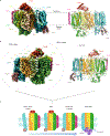
Fig. 1 |
Fig. 1 |. Cryo-EM map of a PSII assembly intermediate (PSII-I) from T. elongatus, segmented into subunits.
a, Fifteen PSII subunits and three assembly factors are coloured and labelled (PSII subunits—D1, D2, CP43, CP47, PsbE, PsbF, PsbH, PsbI, PsbK, PsbL, PsbM, PsbT, PsbX, PsbZ and Psb30; assembly factors—Psb27, Psb28 and tsl0063, which we named Psb34) (front view). b, Parts of PSII that originate from the CP43 module (comprised of CP43, Psb27, PsbZ, Psb30 and PsbK) and the RC47 complex are indicated by dashed lines (back view). c, Schematic model of the PSII assembly process starting with the formation of PSII-I from the CP43 module and RC47. Small PSII subunits were omitted for simplicity.
Fig. 2 |
Fig. 2 |. Psb34 binds to RC47 during attachment of the CP43 module.
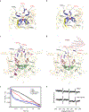
a, Binding site of Psb34 at CP47, close to PsbH (top view), with extended binding of the Psb34 N terminus along the cytoplasmic PSII surface (dashed box). b, Matrix-assisted laser desorption/ionization time-of-flight mass spectrometry (MALDI-TOF MS) analysis of PSII assembly intermediates. Mass spectrum of Psb34 (tsl0063) from the PSII complex (inset) and the fragment spectrum obtained for m/z 5,936.356 with annotated b- and y-ion series matching the Psb34 sequence. Observed fragmentation sites are indicated by dashes in the sequence. Mascot score: 171. c, Subunit composition of Psb34-PSII assembly intermediates analysed by two-dimensional polyacrylamide gel electrophoresis (PAGE). The results are representative of three independent Psb34-TS preparations. a.u., arbitrary units.
Fig. 3 |
Fig. 3 |. The role of the CP47 C terminus in binding of Psb28.
a, Binding of Psb28 at the cytoplasmic/stromal PSII surface (side view, colours correspond to Fig. 1) and continuation of the central Psb28 β-sheet by the CP47 C terminus and the D-E loop of D1 (dashed box). For comparison, mature monomeric PSII (PDB ID 3KZI) is shown in grey. b, Superimposed two-dimensional 1H-15N-HSQC spectra of free Psb28 (blue) and Psb28 bound to the C-terminal peptide of CP47 (magenta). Upper left inset: representation of slow exchange behaviour for the proton amide resonance of T24, ranging from 126.9 to 128.6 ppm in the 15N dimension. c, CSPs of >1 s.d. projected onto the model representation of Psb28. d, Weighted 1H/15N chemical shift perturbations observed for Psb28 on binding to the CP47 peptide. Red line indicates 1 s.d.; residues that yield resonances only in the complex form are indicated in orange. e, Backbone 15N {1H}-heteronuclear NOE of free Psb28 (blue) and Psb28 bound to the C-terminal region of the CP47 peptide (magenta). I/I0 data are presented as mean values ± s.d. derived from n = 3 independent experiments. Smaller I/I0 ratios correspond to regions that exhibit dynamics on the pico- to nanosecond timescale.
Fig. 4 |
Fig. 4 |. Structural changes of the D1 and D2 D-E loops induced by binding of Psb28 and Psb34.
a, Side view of the CP43 antenna protein in PSII-I (teal) and the PSII-M control (light blue). b, Structural changes between PSII-I and the PSII-M control in the cytoplasmic D2 D-E loop (yellow, PSII-I; blue, PSII-M) and attachment of CP43 (teal, PSII-I; light blue, PSII-M control) (top view). Details of the structural changes in the D2 loop are shown in Supplementary Fig. 5a,b. c,d, Side view (c) and top view (d) of the PSII-I structure (orange) compared to the PSII-M control (light blue) and mature monomeric PSII (light red, PDB ID 3KZI). e,f, Side view (e) and top view (f) of the Psb28-induced structural changes in the D1 D-E loop (orange) and perturbation of the QB binding site compared to PSII-M (light blue), which lacks the assembly factors. QA is shown in yellow (PSII-I) or light blue (PSII-M), respectively. See Supplementary Fig. 5c–h for enlarged views of the QA and QB binding sites and the adjacent non-haem iron.
Fig. 5 |
Fig. 5 |. Binding of Psb28 displaces bicarbonate as a ligand of the non-haem iron and protects PSII from damage.
a, The electron transfer from QA to QB is coordinated by the non-haem iron (Fe2+), with the binding of bicarbonate (Bic) serving as a regulatory mechanism in mature PSII (PDB ID 3WU2). b, Binding of Psb28 to the PSII-I assembly intermediate induces a conformational change in the cytoplasmic D2 D-E loop, where the side chain of Glu241 replaces bicarbonate as a ligand of the non-haem iron. The respective fits of the non-haem iron binding sites are shown in (Supplementary Fig. 5e,f). A similar coordination is also found in non-oxygenic bacterial reaction centres (Supplementary Fig. 6c). c, Electron transfer (purple arrows) in mature PSII. Light-induced charge separation at the RC chlorophylls (PD1, PD2, ChlD1 and ChlD2) leads to electron transfer via pheophytin (PheoD1) and plastoquinone A (QA) towards QB. The electron gap at the RC is filled by the OEC. d, Reoxidation of QA by direct and safe charge recombination is favoured in the PSII assembly intermediate, as indicated by the purple arrows. e, Flash-induced fluorescence decay of PSII. Blue lines represent active PSII and red lines correspond to PSII-I. Black and grey lines represent PSII control samples without a functional OEC (Apo-PSII, hydroxylamine-treated PSII; PSII (-OUV), extrinsic proteins removed). Full lines represent mean values calculated from three independent experiments (n = 3), with dotted corridors depicting s.d. f, The protective role of Psb28 binding was further confirmed by electron paramagnetic resonance (EPR) spectroscopy using the spin probe TEMPD, which is specific for 1O2, the major reactive oxygen species in PSII generated by triplet chlorophyll (3P).
Fig. 6 |
Fig. 6 |. The role of Psb27 in Mn4CaO5 cluster assembly.
a, Bottom view of the luminal PSII surface for PSII-I (orange), the PSII-M control (light blue) and mature monomeric PSII (PDB ID 3KZI) (light red). b, Side view of CP43 (teal) and Psb27 (brown) in PSII-I, as well as of CP43 (blue) and PsbO (purple) in mature monomeric PSII (PDB ID 3KZI). Dashed box: CP43 E loop with residues Arg345 and Glu342 (shown as sticks), which form the second coordination sphere of the Mn4CaO5 cluster. We changed the numbering of CP43 residues due to a corrected N-terminal sequence (www.UniProt.org). The residues correspond to Arg357 and Glu354 in previous publications. The high-resolution structure of the Mn4CaO5 cluster is taken from Umena et al. (PDB ID 3WU2). c, Position of the D1 C terminus in PSII-I (orange) and mature monomeric PSII (PDB ID 3KZI) (light red).
Fig. 7 |
Fig. 7 |. Conformational changes within the active site of the Mn4CaO5 cluster. The Mn4CaO5 cluster performs the unique water-splitting reaction of PSII.
a, The active site of the Mn4CaO5 cluster is resolved within our PSII-I structural model but is not yet oxygen evolving. b, Crystal structure of the oxygen evolving, mature PSII (PDB ID 3WU2, resolution 1.9 Å). c, Overlay of both structures, illustrating notable differences in the backbone conformation of the D1 and D2 C-terminal tails. d, Accompanying side-chain rearrangements of the D1 C-terminus. eg, The Cl (e), ionn+ (f) and Mn4CaO5 (g) cluster coordination partners are compared in detail. The validation of the fit to the density of structural details shown here is provided in Supplementary Fig. 7.

Similar articles

Cited by

References

    1. Hohmann-Marriott MF & Blankenship RE Evolution of photosynthesis. Annu. Rev. Plant Biol 62, 515–548 (2011). - PubMed
    1. Sanchez-Baracaldo P & Cardona T On the origin of oxygenic photosynthesis and cyanobacteria. New Phytol 225, 1440–1446 (2020). - PubMed
    1. Vinyard DJ, Ananyev GM & Dismukes GC Photosystem II: the reaction center of oxygenic photosynthesis. Annu. Rev. Biochem 82, 577–606 (2013). - PubMed
    1. Boekema EJ et al. Supramolecular structure of the photosystem II complex from green plants and cyanobacteria. Proc. Natl Acad. Sci. USA 92, 175–179 (1995). - PMC - PubMed
    1. Cox N, Pantazis DA & Lubitz W Current understanding of the mechanism of water oxidation in photosystem II and its relation to XFEL data. Annu. Rev. Biochem 89, 795–820 (2020). - PubMed

MeSH terms

Substances

Supplementary concepts