. 2021 Apr;3(4):513-522.
doi: 10.1038/s42255-021-00366-y. Epub 2021 Apr 12.

Colchicine acts selectively in the liver to induce hepatokines that inhibit myeloid cell activation

Affiliations

Colchicine acts selectively in the liver to induce hepatokines that inhibit myeloid cell activation

Jui-Hsia Weng et al. Nat Metab. 2021 Apr.

Erratum in

Abstract

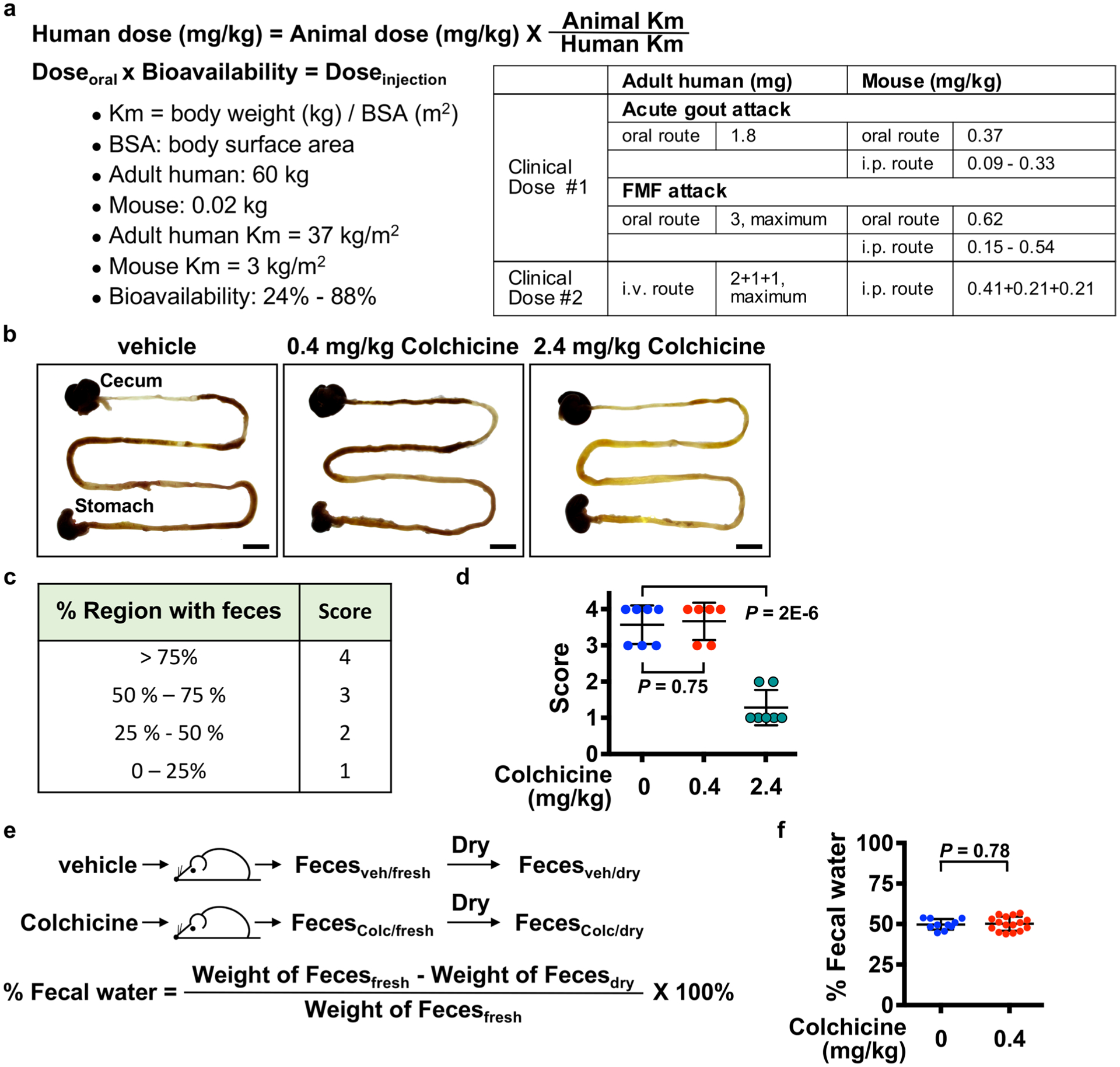
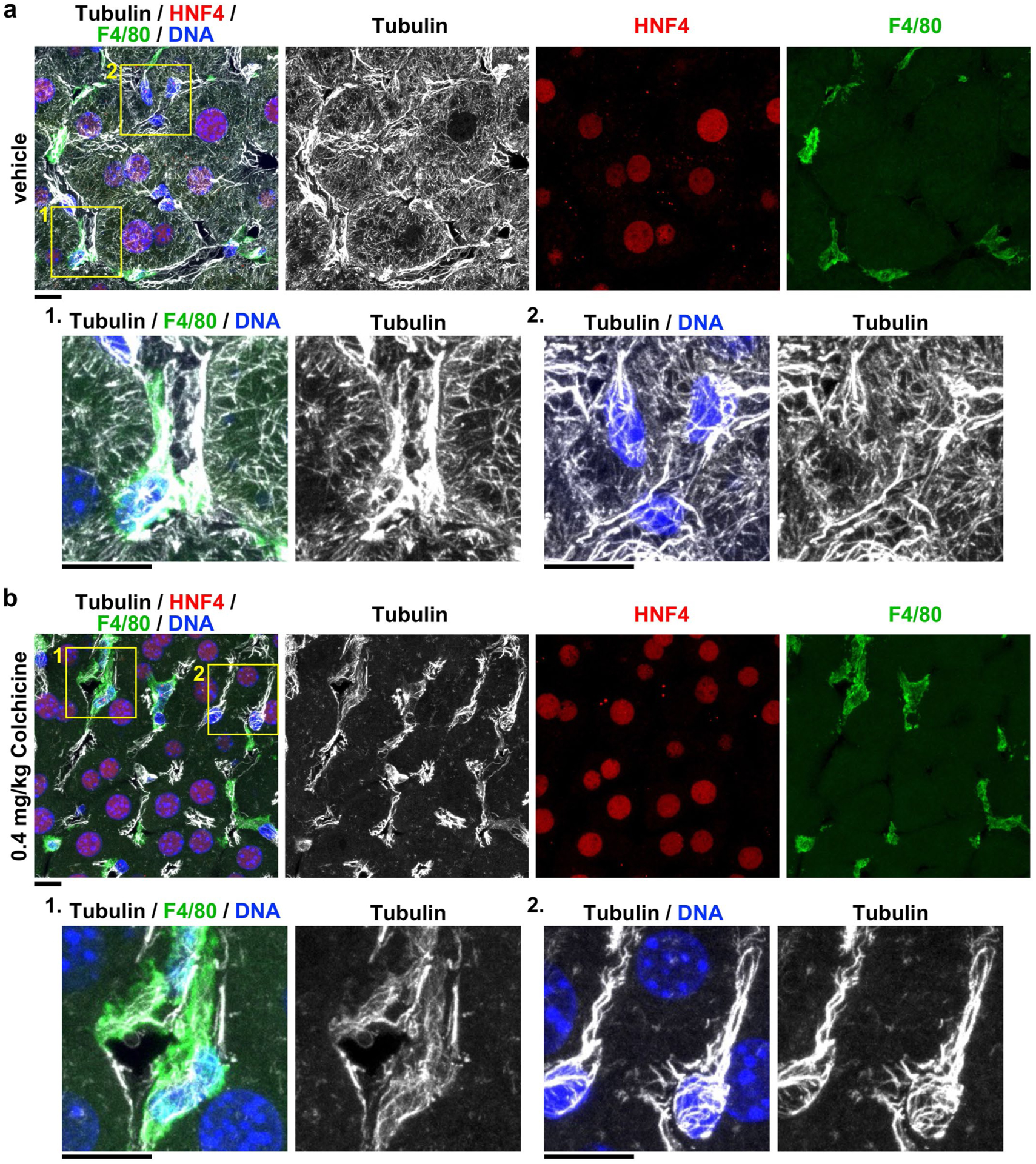
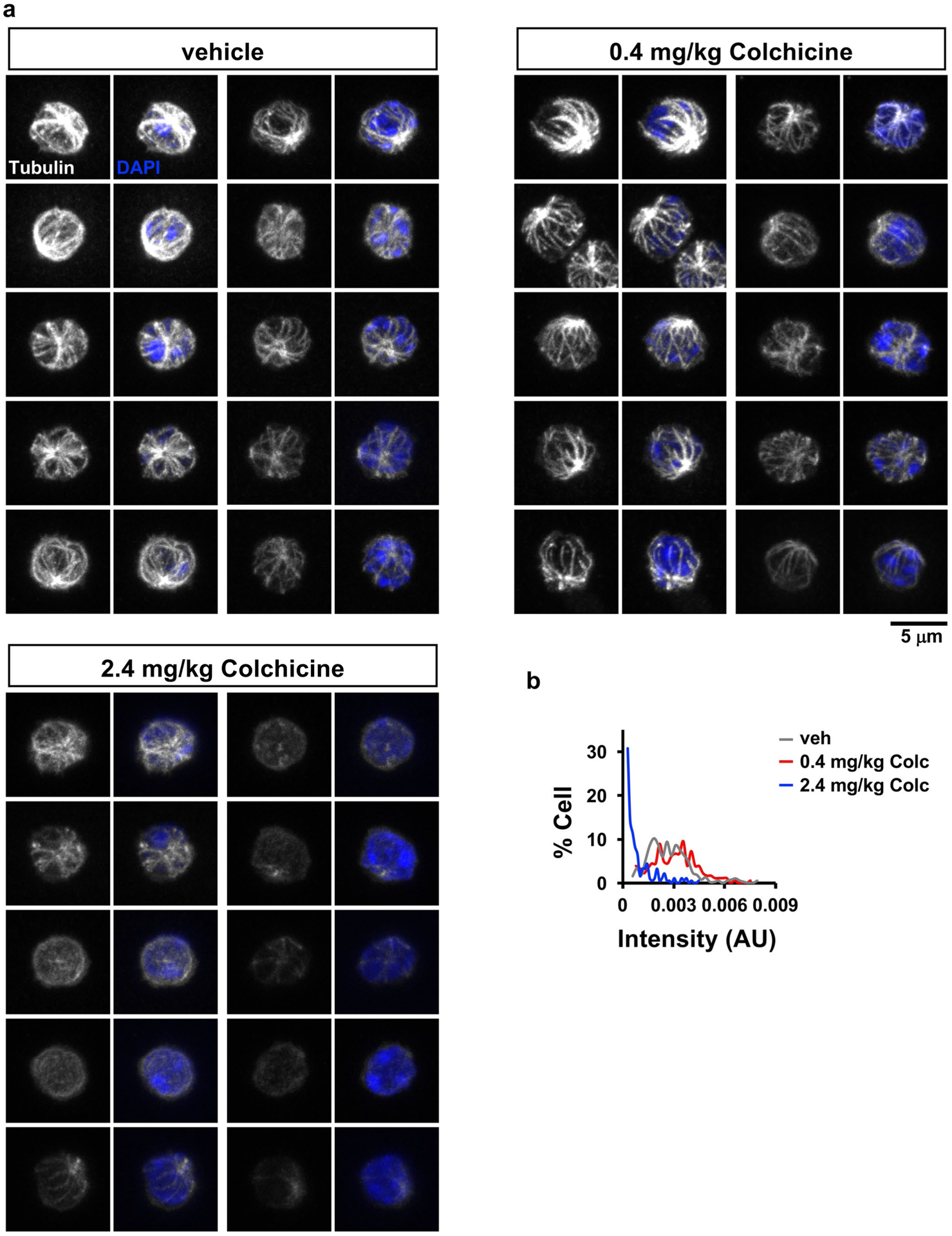
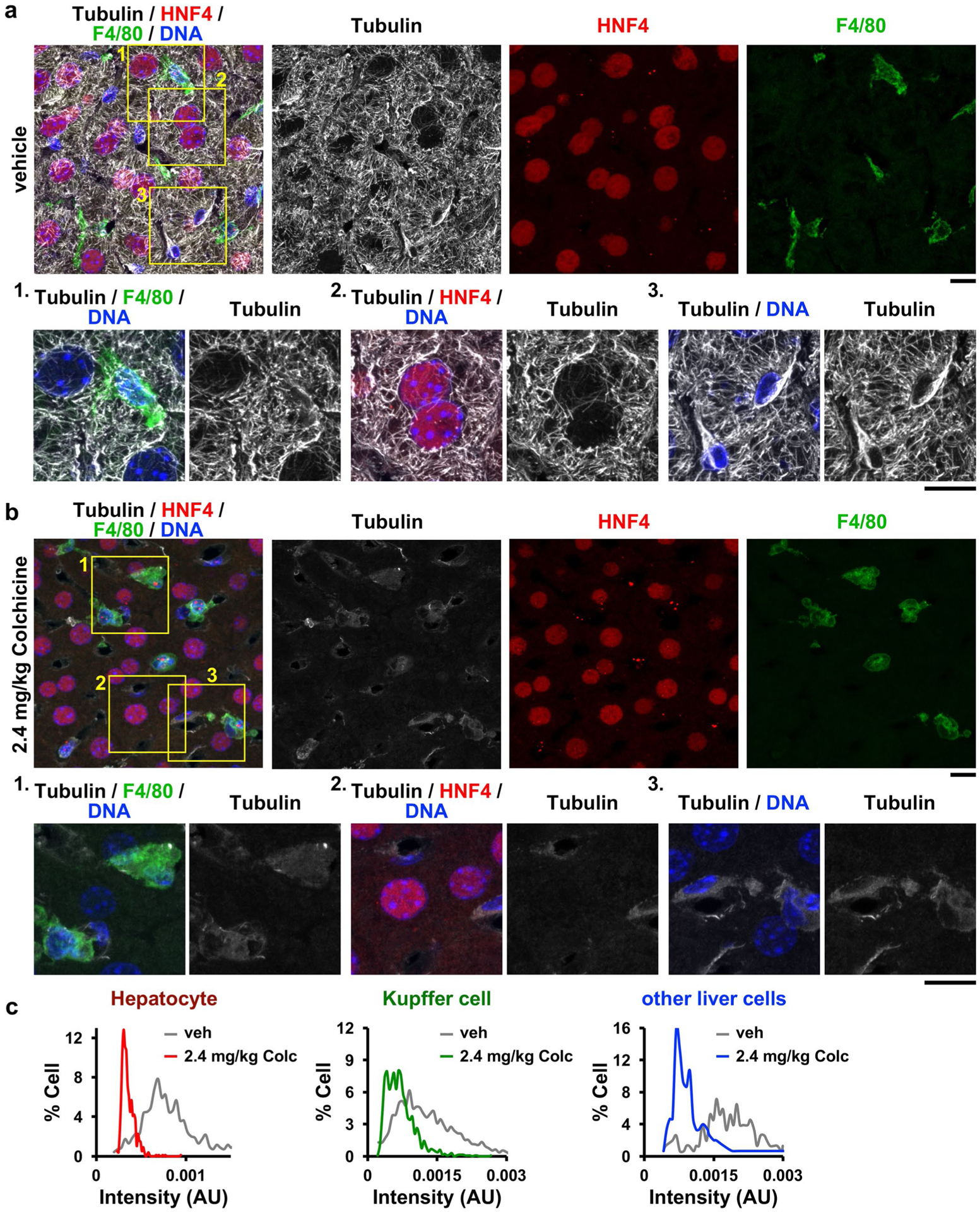
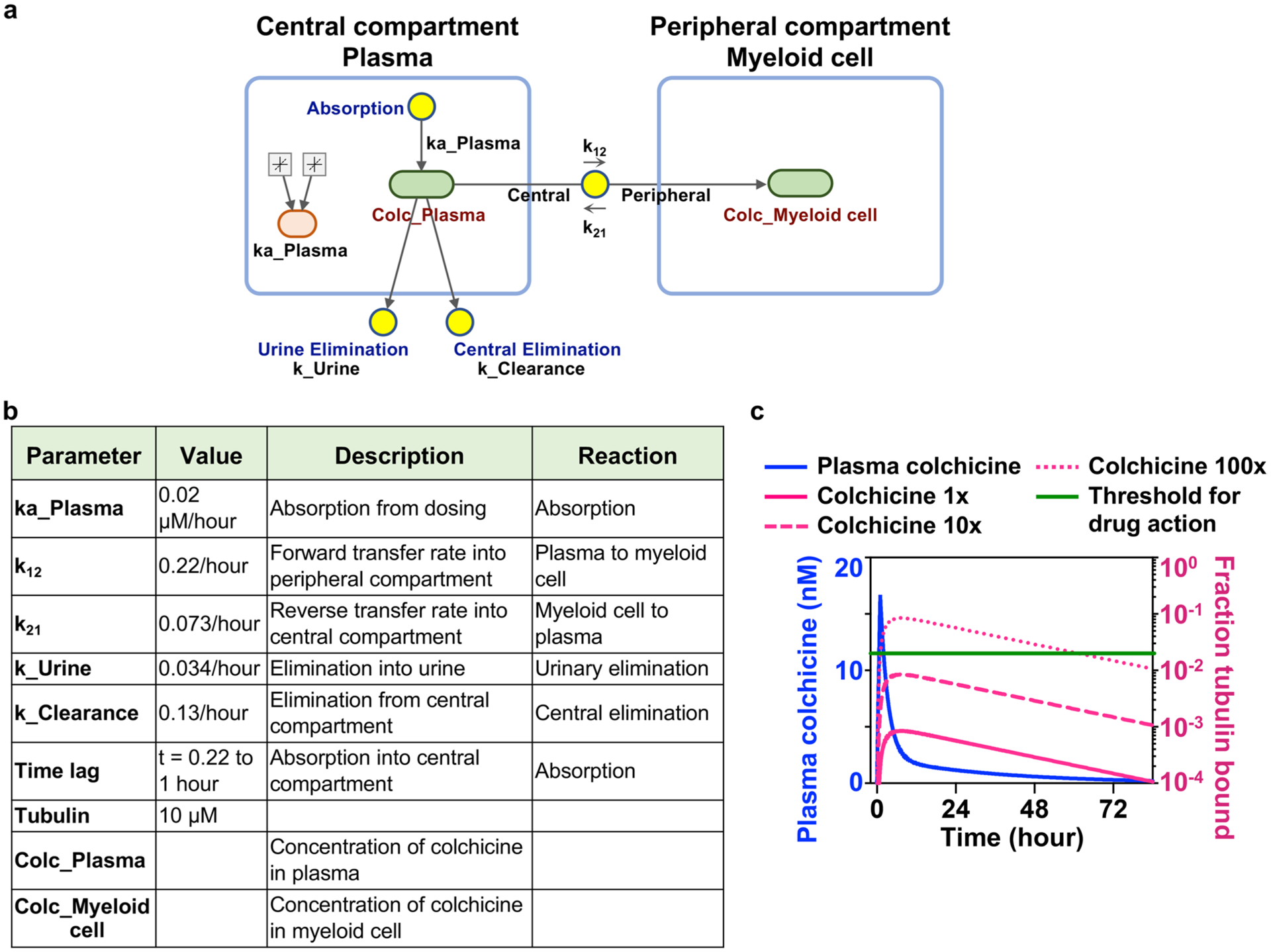
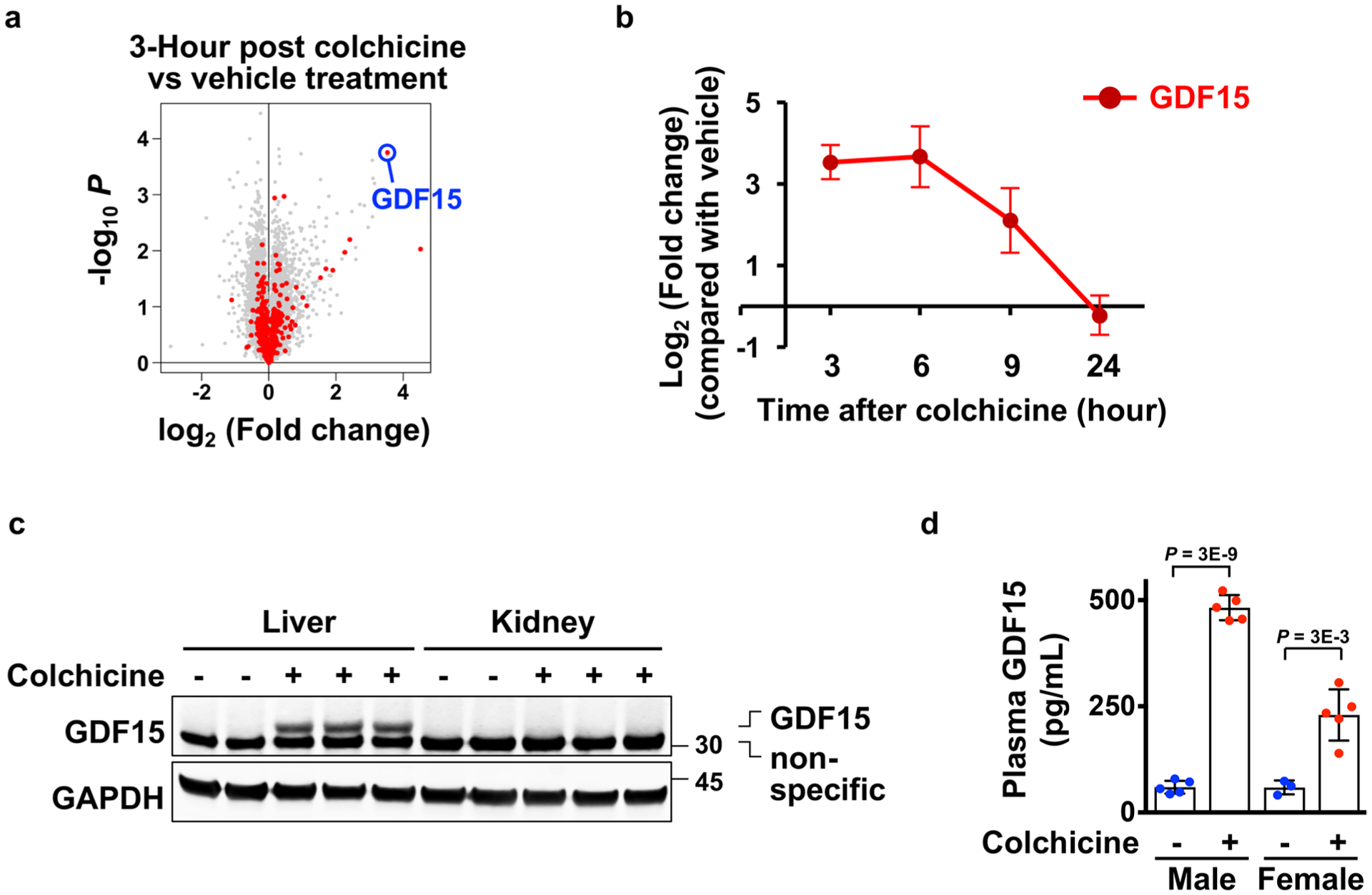
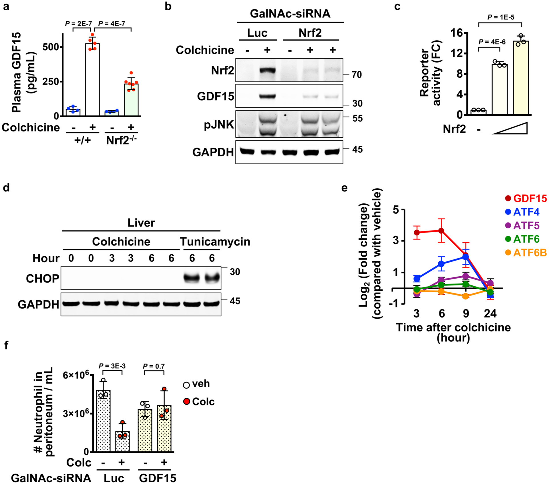
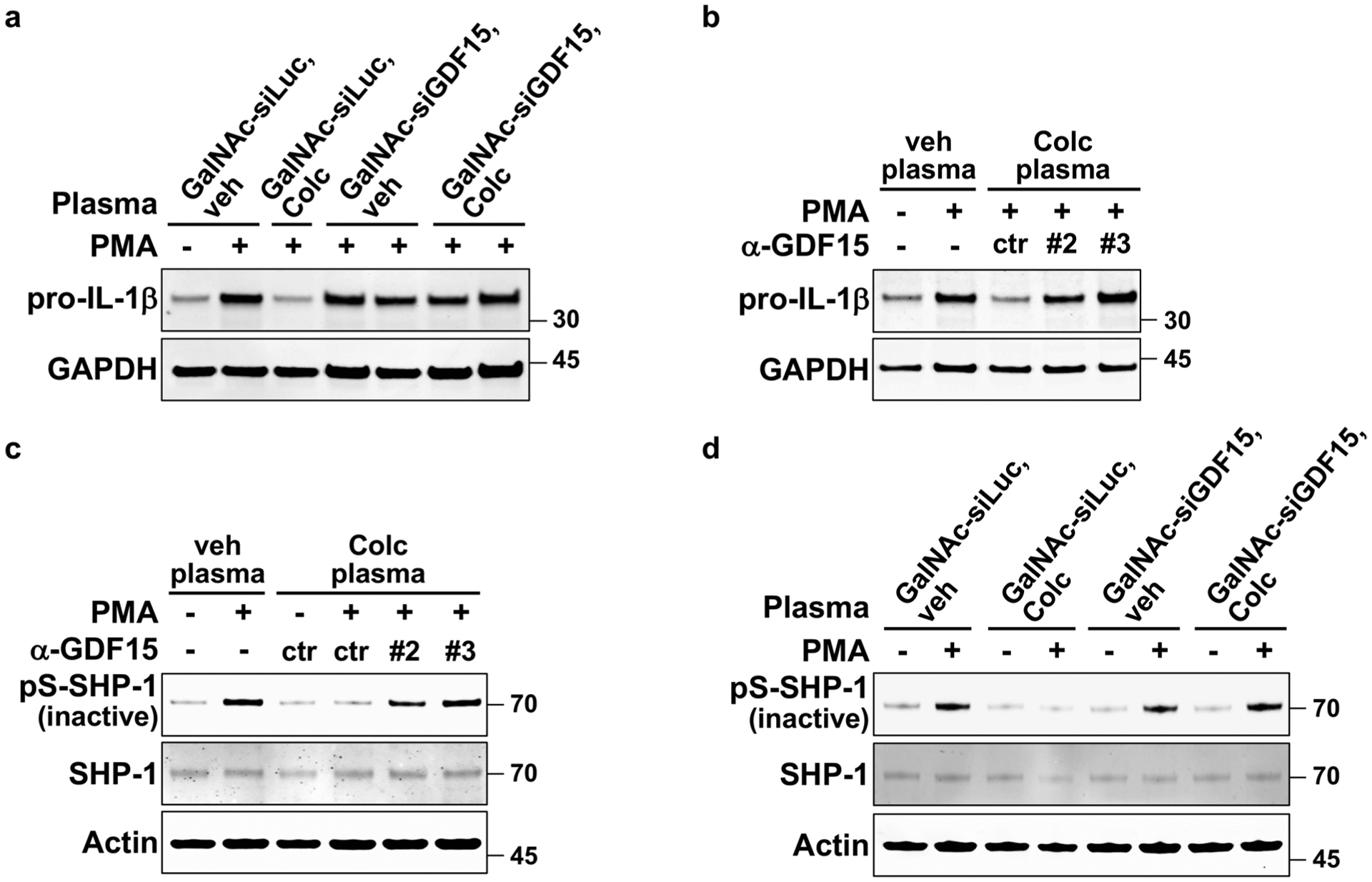
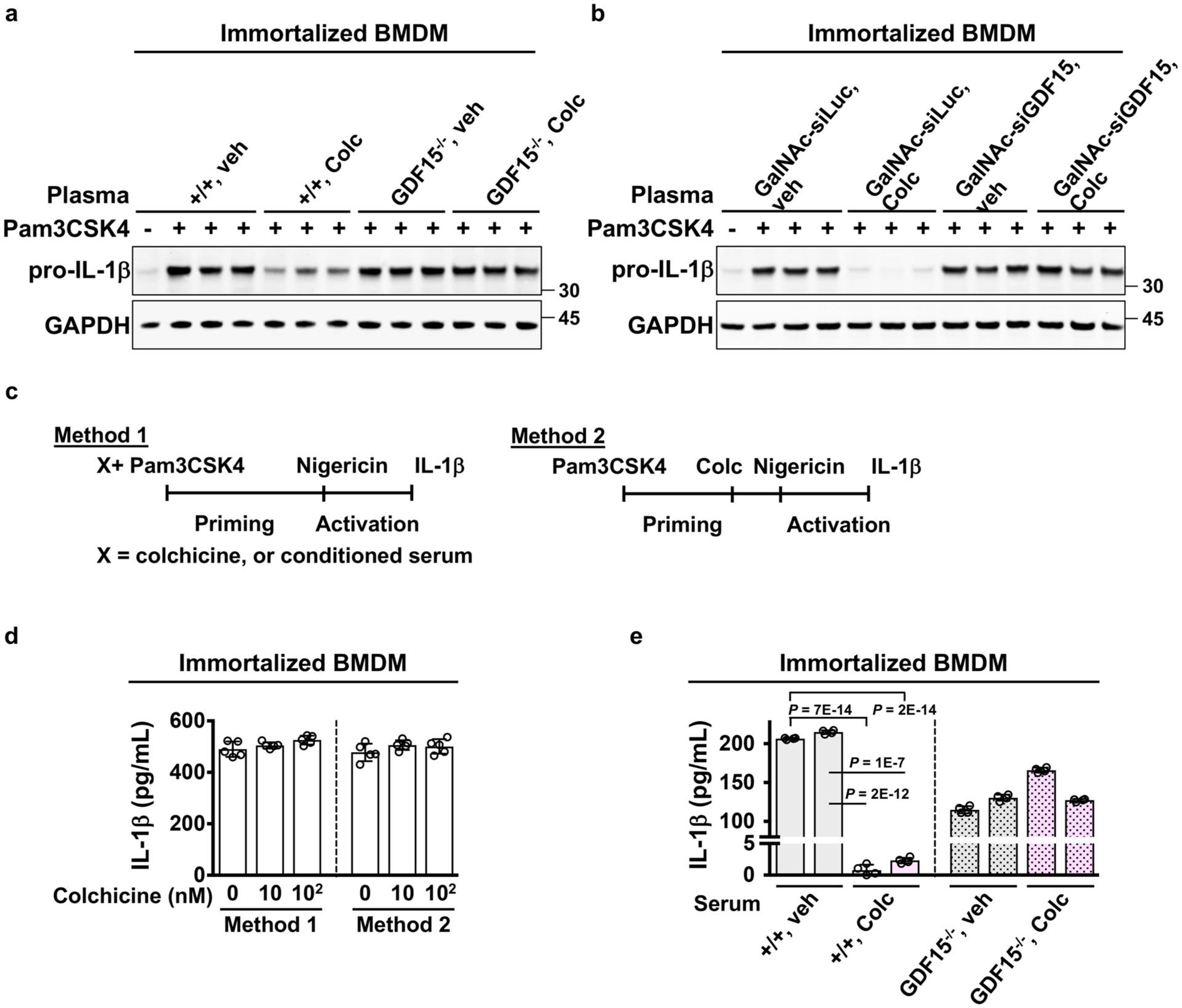
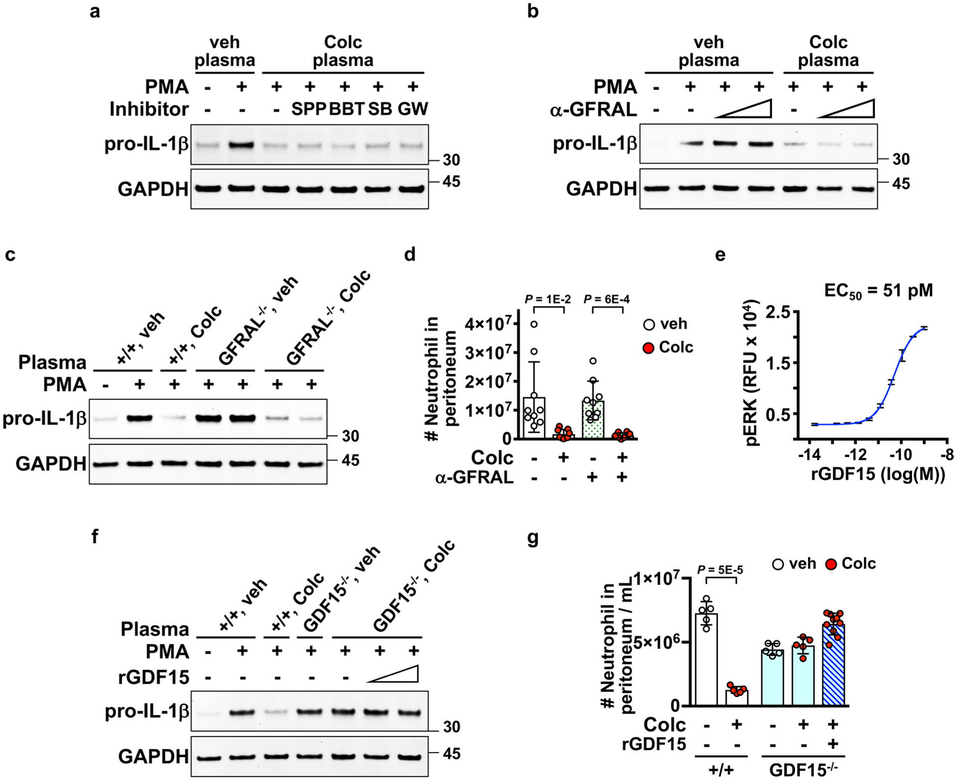
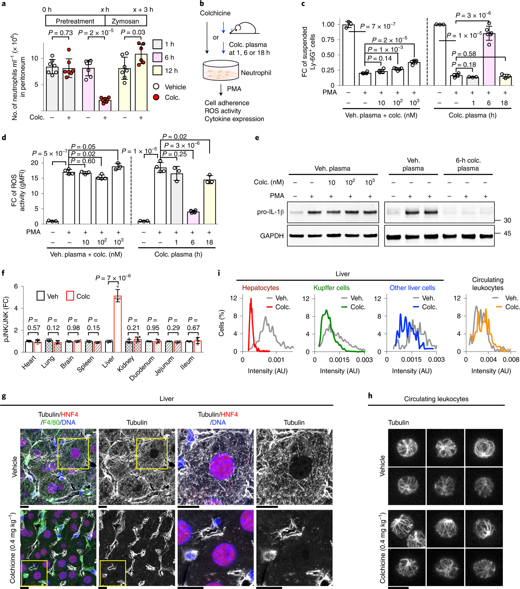
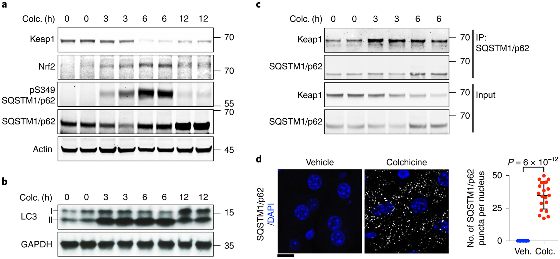
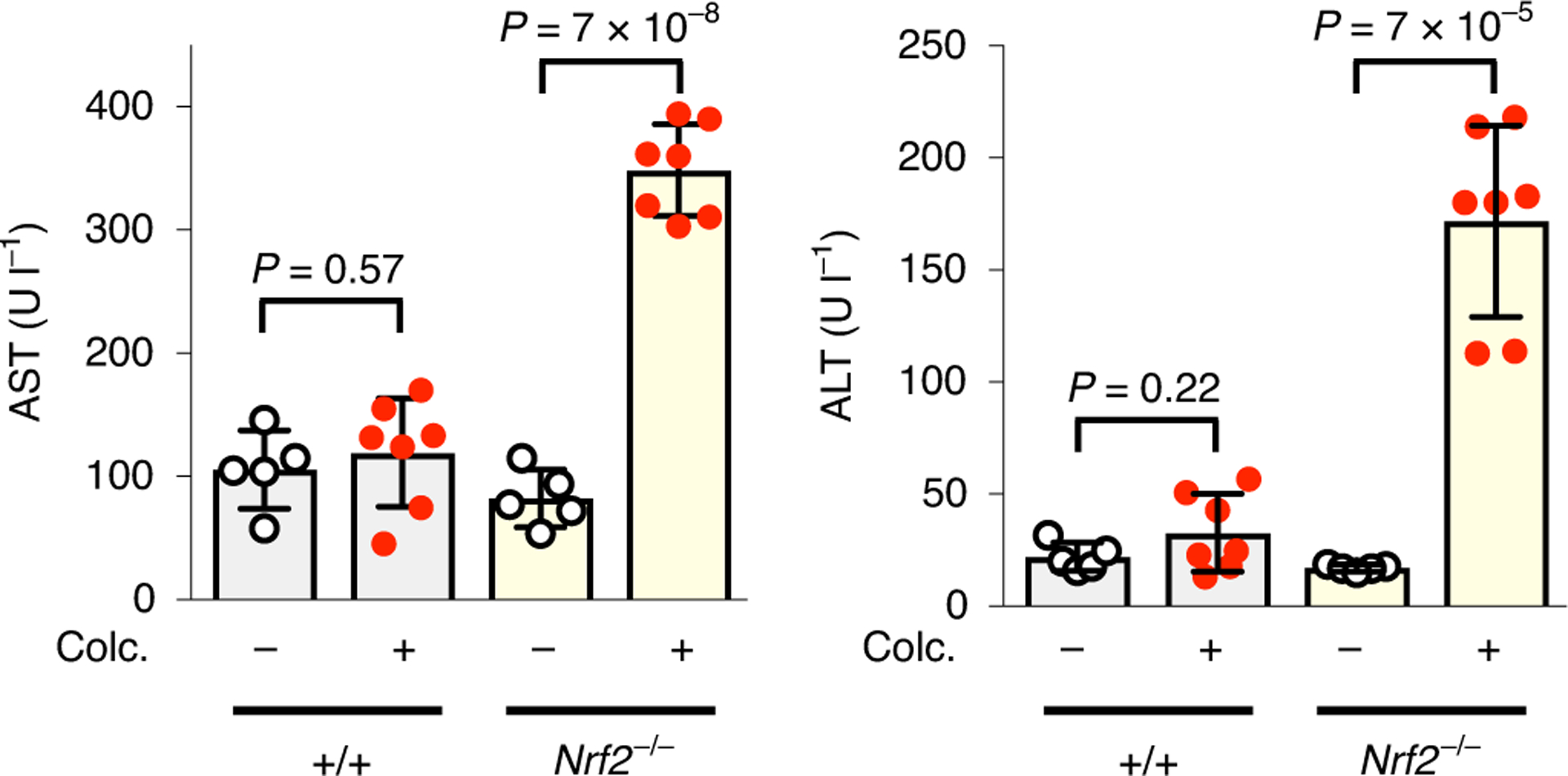
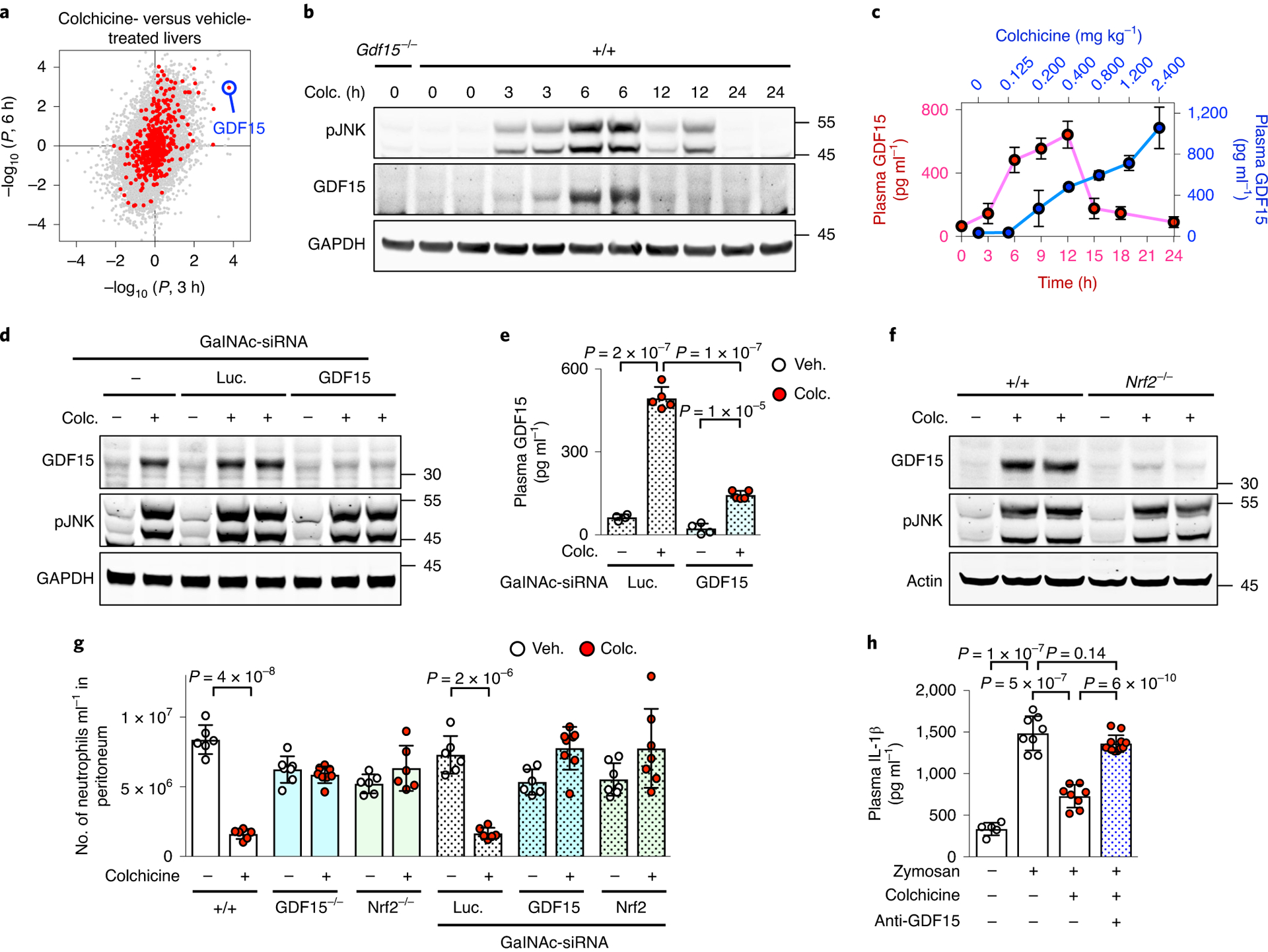
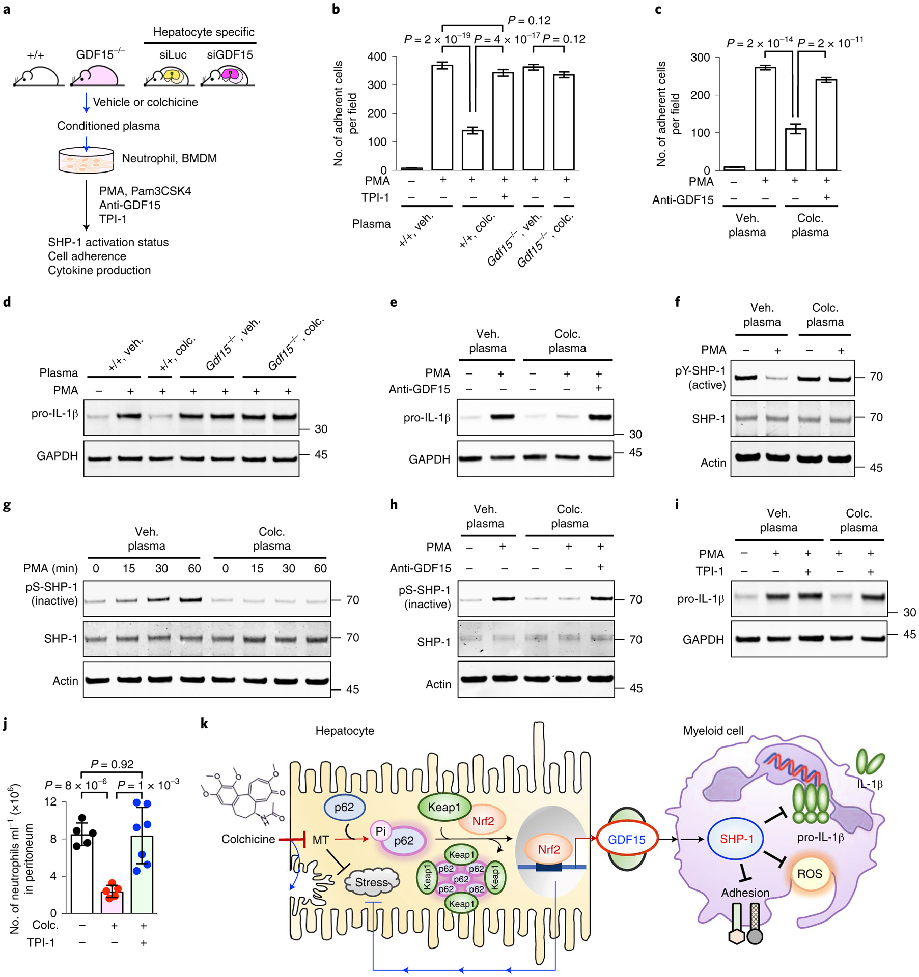
Colchicine has served as a traditional medicine for millennia and remains widely used to treat inflammatory and other disorders. Colchicine binds tubulin and depolymerizes microtubules, but it remains unclear how this mechanism blocks myeloid cell recruitment to inflamed tissues. Here we show that colchicine inhibits myeloid cell activation via an indirect mechanism involving the release of hepatokines. We find that a safe dose of colchicine depolymerizes microtubules selectively in hepatocytes but not in circulating myeloid cells. Mechanistically, colchicine triggers Nrf2 activation in hepatocytes, leading to secretion of anti-inflammatory hepatokines, including growth differentiation factor 15 (GDF15). Nrf2 and GDF15 are required for the anti-inflammatory action of colchicine in vivo. Plasma from colchicine-treated mice inhibits inflammatory signalling in myeloid cells in a GDF15-dependent manner, by positive regulation of SHP-1 (PTPN6) phosphatase, although the precise molecular identities of colchicine-induced GDF15 and its receptor require further characterization. Our work shows that the efficacy and safety of colchicine depend on its selective action on hepatocytes, and reveals a new axis of liver-myeloid cell communication. Plasma GDF15 levels and myeloid cell SHP-1 activity may be useful pharmacodynamic biomarkers of colchicine action.


Conflict of interest statement

Competing interests

H.-C.T. was an employee of Alnylam Pharmaceuticals when the work was done. H.H.L., I.N. and R.V. were employees of NGM Biopharmaceuticals when the work was done. The other authors declare no competing interests.

Figures

Extended Data Fig. 1 |
Extended Data Fig. 1 |. Selection of colchicine doses for mouse treatment.
a, Dose translation from humans to animal studies based on clinical guidelines. The colchicine doses for mouse via the oral or i.p. routes were calculated according to the guidelines from the Food and Drug Administration and the European League Against rheumatism, and were listed in the table. i.p., intraperitoneal. i.v., intravascular. bf, Safety analysis of colchicine based on measurement of gut toxicity. Diarrhea is the dose-limiting toxicity in man. Mice received either vehicle, colchicine at 0.4 mg/kg, or 2.4 mg/kg. b, Intestines were harvested 6 hours after vehicle or colchicine treatment. Scale bars, 1 cm. c, The physiological severity score for diarrhea. d, Colchicine at 0.4 mg/kg did not affect the region of intestine with feces, but 2.4 mg/kg reduced it. Each dot represents one mouse. e, Detection of fecal water content by measuring weight of feces before and after drying. f, Colchicine at 0.4 mg/kg did not affect the fecal water content. Data are represented as mean ± s.d. Two-sided t-tests were used for statistical analysis. A single colchicine dose of 0.4mg/mg i.p. was used in all subsequent experiments unless otherwise indicated.
Extended Data Fig. 2 |
Extended Data Fig. 2 |. Colchicine at a safe dose selectively targets hepatocytes.
Additional images related to Fig. 1g. Microtubules in the livers of mice treated with a, vehicle or b, 0.4 mg/kg colchicine. Livers were collected 6 hours after treatment. Colchicine selectively depolymerized microtubules in hepatocytes, identified by HNF4+. The boxed areas are magnified and shown at the bottom. Liver cells from 4 livers with vehicle or colchicine treatment respectively. Scale bars, 10 μm.
Extended Data Fig. 3 |
Extended Data Fig. 3 |. Colchicine at a safe dose does not damage microtubules in circulating blood cells.
Six hours after treatment of vehicle or colchicine (0.4 mg/kg or 2.4 mg/kg), the whole blood samples were collected and fixed directly. Microtubules were visualized by immunofluorescence and imaged by confocal microscopy. a, representative images of microtubules in circulating blood cells. b, Quantification of microtubule staining intensity. Individual cells exhibited a wide range of microtubule intensities that were similar following vehicle or 0.4 mg/kg colchicine (the safe dose). Microtubules were depolymerized in cells from mice receiving 2.4 mg/kg (the toxic dose). AU, arbitrary unit. Scale bar, 5 μm.
Extended Data Fig. 4 |
Extended Data Fig. 4 |. Colchicine at a toxic dose targets all cell types in the liver.
Microtubules in a, vehicle- or b, 2.4 mg/kg colchicine-treated livers were visualized, and c, intensities of microtubules were quantified. Microtubules were depolymerized in all cell types after treatment of colchicine at 2.4 mg/kg. The boxed areas are magnified and shown at the bottom. AU, arbitrary unit. Scale bars, 10 μm.
Extended Data Fig. 5 |
Extended Data Fig. 5 |. Modeling of human pharmacokinetics-pharmacodynamics (PK-PD) of colchicine.
a, Simulation diagram of two-compartment PK-PD model, based on human pharmacology data from Thomas, G. et al. Two blue boxes represent the central and peripheral compartments, which correspond to plasma and myeloid cell respectively. Light green boxes correspond to reaction species (colchicine concentration in plasma or myeloid cell), and yellow circles correspond to reactions (absorption, urinary elimination, central elimination, and transfer between plasma and myeloid cell). Colchicine absorption takes place from t = 0.22 to t = 1 hour. Tubulin-colchicine binding biochemistry was ignored and all colchicine in the myeloid cells was assumed to bind to tubulin. Fraction bound was calculated based on equations listed in Methods. b, Constants for modeling. c, Human PK-PD modeling of colchicine supported the indirect action of colchicine. The blue line represents colchicine concentrations in plasma over time after colchicine dosing. Magenta lines show the fraction of colchicine-bound tubulin in myeloid cells. Dashed and dotted magenta lines show unbound colchicine concentrated across the cell membrane by a factor of 10 and 100 respectively. The green line marks the 2% threshold of site occupancy for colchicine action.
Extended Data Fig. 6 |
Extended Data Fig. 6 |. GDF15 is a novel hepatokine induced by colchicine.
a, Volcano plot for the gene expression changes of rat livers comparing 3 hours after vehicle to colchicine treatment. Biological triplicate data are from the Open TG-GATEs database. red and gray dots represent genes encoding secreted and non-secreted proteins respectively. GDF15 is highlighted in blue. P, probability value. b, Time course of GDF15 mrNA expression in the rat liver. Data are from Open TG-GATEs. 3 Mice per condition. c, GDF15 was induced in the liver but not kidney collected from colchicine-treated mice. d, ELISA analysis for measurement of plasma GDF15 in both mouse sexes. Induction was reproducibly stronger in males in the C57BL/6J mouse strain. Each dot represents one mouse. 5 Mice per condition. 3 Biological replicates. Data are represented as mean ± s.d. Two-sided t-tests were used for statistical analysis.
Extended Data Fig. 7 |
Extended Data Fig. 7 |. GDF15 is a novel anti-inflammatory hepatokine induced by Nrf2 signaling triggered by colchicine.
a, ELISA analysis for measurement of plasma GDF15 in wild-type and Nrf2 germline knockout (Nrf2−/−) mice. Each dot represents one mouse. b, Immunoblotting analysis revealed that induction of GDF15 in livers was blocked by treatment of hepatocyte-specific sirNA (GalNAc-sirNA) against Nrf2. Luc, luciferase. 3 Biological replicates. c, Activation of GDF15 promoter activity by Nrf2. HEK 293T cells were co-transfected with the luciferase reporter driven by the GDF15 promoter and the Nrf2 plasmid. Luciferase activities were determined from three samples. Data from three independent experiments. d, Immunoblotting analysis for detection of CHOP expression in livers collected at the indicated times after colchicine or Tunicamycin treatment. e, The temporal mrNA expression pattern of the integrated stress response regulators in the rat liver. Data are from Open TG-GATEs. 3 Mice per condition. f, The anti-inflammatory effect of colchicine in the MSU (monosodium urate crystals) peritonitis model depended on GDF15 induction in hepatocytes. Mice were challenged with 1 mg MSU. GalNAc-sirNA, hepatocyte-specific sirNA. Luc, luciferase. Each dot represents one mouse. Data are represented as mean ± s.d. Two-sided t-tests were used for statistical analysis.
Extended Data Fig. 8 |
Extended Data Fig. 8 |. Colchicine-induced GDF15 inhibits pro-IL-1β expression by activating the immuno-inhibitory signal of SHP-1 in primary neutrophils.
Ex vivo activation of neutrophils evaluated by the levels of (ab) pro-IL-1β induction or (cd) phosphorylated SHP-1. Cells were pre-incubated with plasma from the indicated mice and then activated with PMA. a, Induction of GDF15 specifically in hepatocytes was required for the anti-inflammatory activity of post-colchicine plasma, as assayed by pro-IL-1β induction. GalNAc-siLuc, hepatocyte-specific sirNA against luciferase. GalNAc-siGDF15, hepatocyte-specific sirNA against GDF15. b, The anti-inflammatory activity of post-colchicine plasma was blocked by two different clones of antibodies (#2 and #3) against GDF15. ctr, control antibody. veh plasma, plasma from vehicle-treated mice. Colc plasma, plasma from colchicine-treated mice. c, GDF15 neutralization by two clones of anti-GDF15 antibodies (#2 and #3) blocked plasma activity on stabilizing SHP-1 activation. d, Expression of colchicine-induced GDF15 specifically in hepatocytes was required for activating SHP-1. 3 Biological replicates.
Extended Data Fig. 9 |
Extended Data Fig. 9 |. Colchicine-induced GDF15 inhibits pro-IL-1β expression and mature IL-1β secretion from macrophages.
Ex vivo activation of immortalized BMDMs evaluated by the levels of (ab) pro-IL-1β expression and (de) IL-1β secretion. ab, Cells were pre-incubated with plasma from the indicated mice and then activated with Pam3CSK4. Expression of colchicine-induced GDF15 in hepatocytes was required for plasma activity on blocking pro-IL-1β expression. GalNAc-siLuc, hepatocyte-specific sirNA against luciferase. GalNAc-siGDF15, hepatocyte-specific sirNA against GDF15. c, Activation methods for measuring IL-1β production. d, Colchicine did not alter IL-1β production assayed with two activation methods. 5 Samples per condition over 3 biological replicates e, Colchicine-induced GDF15 was required for plasma activity on reducing IL-1β production. 4 Samples per condition over 3 biological replicates Data are represented as mean ± s.d. Two-sided t-tests were used for statistical analysis.
Extended Data Fig. 10 |
Extended Data Fig. 10 |. Canonical mature GDF15-GFRaL-ReT signaling is not involved in the anti-inflammatory actions of colchicine.
ac, and f, Ex vivo activation of neutrophils evaluated by the levels of pro-IL-1β expression. Inhibition of the GFrAL-rET signaling by a, rET inhibitor, SPP86 (SPP) and BBT594 (BTT), or b, a GFrAL neutralizing antibody (α-GFrAL) did not alter post-colchicine plasma activity on blocking pro-IL-1β expression. c, Post-colchicine plasma from wild-type and GFrAL germline knockout mice blocked pro-IL-1β expression. d, GFrAL inhibition by a GFrAL neutralizing antibody did not affect the in vivo anti-inflammatory effect of colchicine in the zymosan peritonitis assay. e, Efficacy of recombinant mature GDF15 (rGDF15) measured by the levels of phosphorylated ErK in a GFrAL-rET reporter cell model. 3 Samples per condition. recombinant GDF15 was a disulfide-linked mature homodimer. recombinant mature GDF15 did not affect f, ex vivo pro-IL-1β expression, and did not inhibit g, in vivo zymosan-induced neutrophil recruitment as colchicine. Each dot represents one mouse. Data are represented as mean ± s.d. Two-sided t-tests were used for statistical analysis.
Fig. 1 |
Fig. 1 |. Colchicine induces anti-inflammatory mediators and selectively targets hepatocytes.
a, Kinetics of in vivo anti-inflammatory effects of colchicine (colc.). At x h (x = 1, 6 or 12) following colchicine treatment, mice were challenged with i.p. zymosan. Three hours later, peritoneal-infiltrated Ly-6G+ neutrophils were detected by FACS analysis. Only 6-h pretreatment of colchicine decreased neutrophil infiltration. Each dot represents one mouse, seven mice per condition. be, Colchicine indirectly inhibited neutrophil activation. b, Ex vivo neutrophil activation assays comparing the effects of colchicine itself, or of plasma from colchicine-treated mice, on inflammation markers in wild-type primary neutrophils following PMA challenge. Direct colchicine treatments were performed with vehicle plasma, so plasma concentrations were the same in all assays. Colchicine plasma (plasma from colchicine-treated mice) was collected at the indicated times after in vivo treatment. c, Neutrophil adhesion to plastic tubes. The y axis indicates cells remaining in suspension. FC, fold change. At least three samples per condition over three biological replicates. Veh. plasma, plasma from vehicle-treated mice. d, rOS generation detected with the rOS-sensitive dye, dihydrorhodamine 123, and measured by FACS analysis. gMFI, geometric mean fluorescence intensity. At least three samples per condition over three biological replicates. e, Pro-IL-1β gene expression by immunoblotting. Colchicine itself lacked activity in all assays, while plasma collected 6 h after colchicine treatment had strong anti-inflammatory activity in all assays. f, In vivo biomarker analysis. Colchicine-activated JNK selectively in the liver. The amounts of phosphorylated and total JNK in nine different tissues from four mice were measured by immunoblotting. Data are represented as mean ± s.d. Two-sided t-tests were used for statistical analysis. gi, Colchicine selectively depolymerized microtubules in hepatocytes. g, Microtubules in hepatocytes (identified by HNF4+), Kupffer cells (identified by F4/80+) and other cell types (HNF4, F4/80 and DAPI+). Images show frozen sections of livers fixed 6 h after vehicle or colchicine treatment, stained and imaged by confocal microscopy. The boxed areas are magnified and shown at the right. Scale bars, 10 μm. h, Microtubules in circulating leukocytes. Scale bar, 5 μm. i, Intensity measurement of microtubules. Approximately 700 hepatocytes, 200 Kupffer cells and 150 other liver cells from four livers following vehicle or colchicine treatment were quantified. AU, arbitrary units.
Fig. 2 |
Fig. 2 |. Colchicine activates Nrf2–Keap1 signalling.
a, Colchicine (colc.) activated the Nrf2–Keap1 pathway in the liver. Immunoblotting analysis was performed with indicated antibodies to identify proteins in this pathway. Colchicine-treated livers were collected at the indicated times. Colchicine triggered degradation of Keap1, stabilization of Nrf2 and phosphorylation of the selective autophagy receptor SQSTM1/p62. b, Colchicine-activated LC3 as measured by conversion of the cytosolic LC3-I isoform to the membrane-bound lapidated LC3-II isoform. c, Colchicine promoted physical interaction between SQSTM1/p62 and Keap1 as measured by co-immunoprecipitation analysis. d, SQSTM1/p62 puncta in the liver from mice with vehicle (veh.) or colchicine treatment were detected by immunofluorescence analysis. The number of SQSTM1/p62 puncta per nucleus was measured. Data are represented as mean ± s.d. Two-sided t-tests were used for statistical analysis; three biological replicates. Scale bar, 10 μm.
Fig. 3 |
Fig. 3 |. Nrf2 protects the liver against colchicine.
Colchicine (colc.)-induced liver toxicity was prevented by Nrf2. Wild-type and Nrf2−/− mice were treated with either vehicle or a safe dose of colchicine as indicated. Serum levels of AST (left) and ALT (right) were measured as standard clinical biomarkers to infer hepatocyte cell death. Each dot represents one mouse; five and seven mice were used for vehicle and colchicine treatment, respectively. Data are presented as mean ± s.d. Two-sided t-tests were used for statistical analysis.
Fig. 4 |
Fig. 4 |. Colchicine-induced GDF15 is an anti-inflammatory hepatokine.
a, Gene expression profile of rat livers comparing vehicle to colchicine (colc.) treatment for 3 h (x axis) and 6 h (y axis). Each dot represents one gene. red and grey dots denote genes encoding secreted and non-secreted proteins respectively, and GDF15 is highlighted in blue. Data are from the Open TG-GATEs toxicogenomic database. All data points were derived from biological triplicates. b, Induction of GDF15 in mouse livers as detected by immunoblotting. Livers were collected at the indicated times. c, Plasma GDF15 as measured by ELISA analysis. red dots show induction kinetics in mice treated with 0.4 mg kg−1 colchicine; blue dots show the dose response after 6-h treatment; five and three mice were used for induction kinetics and dose response, respectively. d,e, Colchicine-induced GDF15 was mainly secreted by hepatocytes. d, Immunoblotting analysis revealed that induction of GDF15 in livers was blocked by treatment of hepatocyte-specific sirNA (GalNAc-sirNA) against GDF15. Luc, luciferase. e, ELISA analysis showed that hepatocyte-specific knockdown of GDF15 reduced colchicine-induced GDF15 in plasma. remaining colchicine-induced plasma GDF15 was probably derived from the liver due to incomplete knockdown. At least four mice were used per condition. f, Nrf2 was required for colchicine-induced GDF15 expression in liver hepatocytes. g,h, In vivo peritonitis induced by i.p. zymosan challenge and assayed by neutrophil recruitment (g) and plasma IL-1β levels (h). Both Nrf2 and GDF15 were required for in vivo anti-inflammatory activity of colchicine using germline knockouts, hepatocyte-specific sirNA knockdowns and a GDF15-neutralizing antibody (anti-GDF15). Each dot represents one mouse, at least six mice per condition and three biological replicates. Data are represented as mean ± s.d. Two-sided t-tests were used for statistical analysis. Veh., vehicle.
Fig. 5 |
Fig. 5 |. SHP-1 phosphatase mediates the anti-inflammatory effects of colchicine-induced hepatokines on myeloid cells.
a, Ex vivo myeloid cell activation assays. Neutrophils from untreated mice or immortalized BMDMs were incubated in plasma collected from mice (wild type, Gdf15−/− or hepatocyte-specific sirNA knockdowns) following vehicle (veh.) or colchicine (colc.) treatment, then challenged with PMA (neutrophils) or Pam3CSK4 (BMDMs). Anti-GDF15, GDF15-neutralizing antibody; TPI-1, a SHP-1 inhibitor. b, PMA-stimulated adhesion of neutrophils to coverslips was inhibited by plasma from colchicine-treated wild-type mice, but not by Gdf15−/−. Co-treatment with TPI-1 blocked the anti-adhesive activity of post-colchicine plasma. c, Inhibition of GDF15 using a GDF15-neutralizing antibody blocked the anti-inflammatory activity of post-colchicine plasma. Veh. plasma, plasma from vehicle-treated mice; colc. plasma, plasma from colchicine-treated mice. Two-sided t-tests were used for statistical analysis. d, Induction of pro-IL-1β was blocked by post-colchicine plasma from wild-type mice, but not from Gdf15−/− mice. e, Co-treatment with a GDF15-neutralizing antibody blocked plasma activity. fh, SHP-1 was activated by colchicine-induced GDF15. Phosphorylation of SHP-1 at Y564 marks the active form of SHP-1, while phosphorylation at S591 marks the inactive form. i, TPI-1 rescued pro-IL-1β induction, which was blocked by post-colchicine plasma. Post-colchicine plasma increased the active form (f) and decreased the inactive form (g) of SHP-1. h, The anti-inflammatory activity of post-colchicine plasma was blocked by inhibition of GDF15. j, SHP-1 inhibition by TPI-1 blocked the in vivo anti-inflammatory effect of colchicine in the zymosan peritonitis assay. Each dot represents one mouse. Data are presented as mean ± s.d. k, Model for indirect anti-inflammatory action of colchicine. Colchicine activates Nrf2–Keap1 signalling in hepatocytes, which protects them from damage and induction of a novel anti-inflammatory hepatokine, GDF15, into plasma. Colchicine-induced GDF15 then acts on circulating myeloid cells by supporting the active state of SHP-1.

References

    1. Medzhitov R Origin and physiological roles of inflammation. Nature 454, 428–435 (2008). - PubMed
    1. Dinarello CA Anti-inflammatory agents: present and future. Cell 140, 935–950 (2010). - PMC - PubMed
    1. Dasgeb B et al. Colchicine: an ancient drug with novel applications. Br. J. Dermatol 178, 350–356 (2018). - PMC - PubMed
    1. Nidorf SM, Eikelboom JW, Budgeon CA & Thompson PL Low-dose colchicine for secondary prevention of cardiovascular disease. J. Am. Coll. Cardiol 61, 404–410 (2013). - PubMed
    1. Tardif JC et al. Efficacy and safety of low-dose colchicine after myocardial infarction. N. Engl. J. Med 381, 2497–2505 (2019). - PubMed

Publication types

MeSH terms

LinkOut - more resources