. 2021 Jun;23(6):437.
doi: 10.3892/mmr.2021.12076. Epub 2021 Apr 13.

Astragaloside IV alleviates liver injury in type 2 diabetes due to promotion of AMPK/mTOR‑mediated autophagy

Affiliations

Astragaloside IV alleviates liver injury in type 2 diabetes due to promotion of AMPK/mTOR‑mediated autophagy

Yunfeng Zhu et al. Mol Med Rep. 2021 Jun.

Abstract

Diabetic liver injury is a serious complication of type 2 diabetes mellitus (T2DM), which is often irreversible in the later stage, and affects the quality of life. Autophagy serves an important role in the occurrence and development of diabetic liver injury. For example, it can improve insulin resistance (IR), dyslipidaemia, oxidative stress and inflammation. Astragaloside IV (AS‑IV) is a natural saponin isolated from the plant Astragalus membranaceus, which has comprehensive pharmacological effects, such as anti‑oxidation, anti‑inflammation and anti‑apoptosis properties, as well as can enhance immunity. However, whether AS‑IV can alleviate diabetic liver injury in T2DM and its underlying mechanisms remain unknown. The present study used high‑fat diets combined with low‑dose streptozotocin to induce a diabetic liver injury model in T2DM rats to investigate whether AS‑IV could alleviate diabetic liver injury and to identify its underlying mechanisms. The results demonstrated that AS‑IV treatment could restore changes in food intake, water intake, urine volume and body weight, as well as improve liver function and glucose homeostasis in T2DM rats. Moreover, AS‑IV treatment promoted suppressed autophagy in the liver of T2DM rats and improved IR, dyslipidaemia, oxidative stress and inflammation. In addition, AS‑IV activated adenosine monophosphate‑activated protein kinase (AMPK), which inhibited mTOR. Taken together, the present study suggested that AS‑IV alleviated diabetic liver injury in T2DM rats, and its mechanism may be associated with the promotion of AMPK/mTOR‑mediated autophagy, which further improved IR, dyslipidaemia, oxidative stress and inflammation. Thus, the regulation of autophagy may be an effective strategy to treat diabetic liver injury in T2DM.

Keywords: AS‑IV; diabetic liver injury; autophagy; AMPK; mTOR.


Conflict of interest statement

The authors declare that they have no competing interests.

Figures

Figure 1.
Figure 1.
Effects of AS-IV on food intake, water intake, urine volume and body weight in T2DM rats. The (A) 24 h food intake, (B) 24 h water intake and (C) 24 h urine volume at the end of the 4 and 8th weeks after administration. (D) Body weight from 0–16 weeks (the administration was started on the 9th week). Data are presented as the mean ± SD, n=6. ***P<0.001 vs. control group; #P<0.05, ##P<0.01 vs. model group; &P<0.05 vs. 4th week. Con, Control; Mod, Model; AS-IV, Astragaloside IV (80 mg/kg); Met, Metformin (200 mg/kg).
Figure 2.
Figure 2.
AS-IV attenuates liver function in T2DM rats. (A) H&E staining of liver tissues (magnification, ×400). (B) Masson staining of liver tissues (magnification, ×400). (C) Quantitative analysis of positive areas of Masson staining was normalised to the normal control group. (D) AST and (E) ALT levels in serum. (F) Ratio of liver weight to body weight. Data are presented as the mean ± SD, n=6. ***P<0.001 vs. control group; #P<0.05, ##P<0.01, ###P<0.001 vs. model group. Con, Control; Mod, Model; AS-IV, Astragaloside IV (80 mg/kg); Met, Metformin (200 mg/kg); AST, aspartate aminotransferase; ALT, alanine aminotransferase.
Figure 3.
Figure 3.
AS-IV maintains glucose homeostasis in T2DM rats. (A) FBG and (B) FINS levels in serum. (C) HOMA-IR. (D) GSP levels in serum. (E) FBG levels from 0–8 weeks after intervention. (F) Oral glucose tolerance test results and AUC. Data are presented as the mean ± SD, n=6. ***P<0.001 vs. control group; #P<0.05, ##P<0.01, ###P<0.001 vs. model group; &P<0.05, &&P<0.01, &&&P<0.001 vs. week 0. Con, Control; Mod, Model; AS-IV, Astragaloside IV (80 mg/kg); Met, Metformin (200 mg/kg); FBG, fasting blood glucose; GSP, glycosylated serum protein; FINS, fasting insulin; HOMA-IR, homeostasis model assessment of insulin resistance; AUC, area under the curve.
Figure 4.
Figure 4.
AS-IV improves dyslipidaemia and liver lipid deposition in T2DM rats. (A) Oil red O staining of liver tissues (magnification, ×400). (B) Quantitative analysis of positive areas of Oil red O staining was normalised to the normal control group. (C) TG, (D) TC, (E) HDL-C and (F) LDL-C levels in serum. Data are presented as the mean ± SD, n=3-6. ***P<0.001 vs. control group; #P<0.05, ###P<0.001 vs. model group. Con, Control; Mod, Model; AS-IV, Astragaloside IV (80 mg/kg); Met, Metformin (200 mg/kg); TC, total cholesterol; TG, triglyceride; HDL-C, high-density lipoprotein cholesterol; LDL-C, low-density lipoprotein cholesterol.
Figure 5.
Figure 5.
AS-IV ameliorates inflammation and oxidative stress in the liver of T2DM rats. (A) TNF-α and (B) IL-6 levels in serum. (C) Protein expression levels of TNF-α, IL-6 and HO1 in the liver tissues were detected via western blotting. Semi-quantitative analysis of (D) TNF-α, (E) IL-6 and (F) HO-1 was normalised to β-actin. Data are presented as the mean ± SD, n=3. ***P<0.001 vs. control group; #P<0.05, ##P<0.01, ###P<0.001 vs. model group. Con, Control; Mod, Model; AS-IV, Astragaloside IV (80 mg/kg); Met, Metformin (200 mg/kg); HO-1, heme oxygenase-1.
Figure 6.
Figure 6.
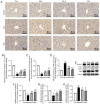
AS-IV activates the suppressed autophagy in the liver of T2DM rats. (A) Representative images of Beclin1, LC3 and P62 (immunohistochemical staining; magnification, ×400). Quantitative analysis of (B) Beclin1, (C) LC3 and (D) P62 were normalized to the normal control group. (E) Protein expression levels of Beclin1, LC3 and P62 in the liver tissues were detected via western blotting. Semi-quantitative analysis of (F) Beclin1, (G) LC3 and (H) P62 was normalised to β-actin. Data are presented as the mean ± SD, n=3. *P<0.05, ***P<0.001 vs. control group; #P<0.05, ##P<0.01, ###P<0.001 vs. model group. Con, Control; Mod, Model; AS-IV, Astragaloside IV (80 mg/kg); Met, Metformin (200 mg/kg).
Figure 7.
Figure 7.
Transmission electron microscopy images showing the ultrastructure of lipids (white arrows) and autophagosomes (black arrows). n=3 for each group. Con, Control; Mod, Model; AS-IV, Astragaloside IV (80 mg/kg); Met, Metformin (200 mg/kg).
Figure 8.
Figure 8.
AS-IV activates autophagy via the AMPK/mTOR pathway in the liver of T2DM rats. (A) Protein expression levels of AMPK, p-AMPK, mTOR and p-mTOR in liver tissues were detected via western blotting. (B and C) Semi-quantitative analysis of p-AMPK/AMPK ratio and p-mTOR/mTOR ratio. Data are presented as the mean ± SD, n=3. **P<0.01, ***P<0.001 vs. control group; #P<0.05, ##P<0.01, ###P<0.001 vs. model group. Con, Control; Mod, Model; AS-IV, Astragaloside IV (80 mg/kg); Met, Metformin (200 mg/kg); p-, phosphorylated; AMPK, adenosine monophosphate-activated protein kinase.

Similar articles

Cited by

References

    1. Tan SY, Mei Wong JL, Sim YJ, Wong SS, Mohamed Elhassan SA, Tan SH, Ling Lim GP, Rong Tay NW, Annan NC, Bhattamisra SK, Candasamy M. Type 1 and 2 diabetes mellitus: A review on current treatment approach and gene therapy as potential intervention. Diabetes Metab Syndr. 2019;13:364–372. doi: 10.1016/j.dsx.2018.10.008. - DOI - PubMed
    1. Zheng Y, Ley SH, Hu FB. Global aetiology and epidemiology of type 2 diabetes mellitus and its complications. Nat Rev Endocrinol. 2018;14:88–98. doi: 10.1038/nrendo.2017.151. - DOI - PubMed
    1. Shima T, Uto H, Ueki K, Takamura T, Kohgo Y, Kawata S, Yasui K, Park H, Nakamura N, Nakatou T, et al. Clinicopathological features of liver injury in patients with type 2 diabetes mellitus and comparative study of histologically proven nonalcoholic fatty liver diseases with or without type 2 diabetes mellitus. J Gastroenterol. 2013;48:515–525. doi: 10.1007/s00535-012-0653-5. - DOI - PubMed
    1. Hsiang JC, Gane EJ, Bai WW, Gerred SJ. Type 2 diabetes: A risk factor for liver mortality and complications in hepatitis B cirrhosis patients. J Gastroenterol Hepatol. 2015;30:591–599. doi: 10.1111/jgh.12790. - DOI - PubMed
    1. Bedi O, Aggarwal S, Trehanpati N, Ramakrishna G, Krishan P. Molecular and pathological events involved in the pathogenesis of diabetes-associated nonalcoholic fatty liver disease. J Clin Exp Hepatol. 2019;9:607–618. doi: 10.1016/j.jceh.2018.10.004. - DOI - PMC - PubMed

MeSH terms