. 2021 Jun;47(6):98.
doi: 10.3892/ijmm.2021.4931. Epub 2021 Apr 13.

Mechanisms of microRNA‑142 in mitochondrial autophagy and hippocampal damage in a rat model of epilepsy

Affiliations

Mechanisms of microRNA‑142 in mitochondrial autophagy and hippocampal damage in a rat model of epilepsy

Du Xiao et al. Int J Mol Med. 2021 Jun.

Abstract

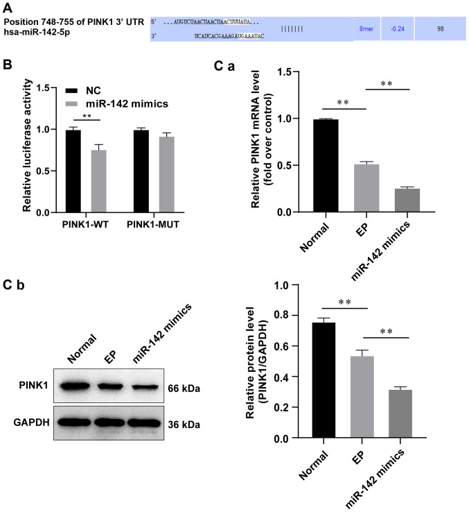
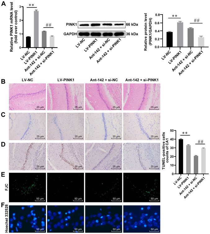
Researchers have confirmed the microRNA (miRNA/miR)‑epilepsy association in rodent models of human epilepsy via a comprehensive database. However, the mechanisms of miR‑142 in epilepsy have not been extensively studied. In the present study, a rat model of epilepsy was first established by an injection of lithium chloride‑pilocarpine and the successful establishment of the model was verified via electroencephalogram monitoring. The levels of miR‑142, phosphatase and tensin homolog deleted on chromosome 10 (PTEN)‑induced putative kinase 1 (PINK1), marker proteins of mitochondrial autophagy, and apoptosis‑related proteins were measured. Additionally, the pathological changes in the hippocampus, the ultrastructure of the mitochondria, and degeneration and the apoptosis of neurons were observed using different staining methods. The malondialdehyde (MDA) content and superoxide dismutase (SOD) activity in the hippocampus, mitochondrial membrane potential (MTP) and reactive oxygen species (ROS) generation were detected. Furthermore, the targeting association between miR‑142 and PINK1 was predicted and verified. Consequently, apoptosis increased, and mitochondrial autophagy decreased, in the hippocampus of epileptic rats. Following miR‑142 inhibition, the epileptic rats exhibited an increased Bax expression, a decreased Bcl‑2 expression, upregulated marker protein levels of mitochondrial autophagy, a reduced MDA content, an enhanced SOD activity, an increased MTP and decreased ROS generation. PINK1 is a target gene of miR‑142, and its overexpression protected against hippocampal damage. Taken together, the results of the present study demonstrated that miR‑142 inhibition promotes mitochondrial autophagy and reduces hippocampal damage in epileptic rats by targeting PINK1. These findings may provide useful information for the treatment of epilepsy.

Keywords: epilepsy; microRNA‑142; PTEN‑induced putative kinase 1; mitochondrial autophagy; hippocampal damage.


Conflict of interest statement

The authors declare that they have no competing interests.

Figures

Figure 1
Figure 1
Successful establishment of the model of epilepsy. (A) EEG result of the cortical electrode in rats; (B) EEG result of the hippocampal electrode in rats. 'Normal' indicates results in normal rats, showing no obvious paroxysmal rhythm and epileptic discharge; 'EP' indicates results in epileptic rats 3 h after the epileptic seizure, showing obvious epileptic discharges, spikes and sharp waves. n=6. EEG, electroencephalogram.
Figure 2
Figure 2
miR-142 inhibition reduces ROS and apoptosis in hippocampal neurons in epileptic rats. (A) Relative miR-142 expression in normal rats and epileptic rats measured by RT-qPCR, n=3. (B and C) Representative images of the histopathological sections of the hippocampus in epileptic rats detected by H&E staining and Nissl staining, n=6. (D-F) Representative images of apoptosis and degeneration in the hippocampal CA3 region of epileptic rats detected by the TUNEL assay, FJC staining and Hoechst 332528 staining, n=6. (G) Representative images of the neuron structure in the hippocampus of epileptic rats observed under TEM, n=3. (H) Relative MDA and SOD contents in normal rats and epileptic rats measured by the TBA and WST-8 methods, n=3. (I) Protein levels of Bcl-2 and Bax in the hippocampus of normal rats and epileptic rats measured by western blot analysis, n=3. The data were analyzed using one-way ANOVA and Tukey's multiple comparisons test as a post hoc test. **P<0.01, compared with the normal group; ##P<0.01, compared with the antagomir-NC group. miR-142, microRNA-142; ROS, reactive oxygen species; RT-qPCR, reverse transcription-quantitative polymerase chain reaction; TUNEL, terminal deoxynucleotidyl transferase (TdT)-mediated dUTP nick end labeling; H&E, hematoxylin and eosin; FJC, Fluoro-Jade C; MDA, malondialdehyde; SOD, superoxide dismutase; TBA, thiobarbituric acid; WST-8, water-soluble tetrazolium salt-8; Bcl-2, B-cell lymphoma-2; Bax, Bcl-2-associated X; ANOVA, analysis of variance.
Figure 3
Figure 3
PINK1 is the target gene of miR-142. (A) Binding sequence of miR-142 and PINK1 was analyzed using bioinformatics software. (B) The targeting association between miR-142 and PINK1 was validated by the dual luciferase report gene assay. The data were analyzed by two-way ANOVA, and the pairwise comparisons following ANOVA were analyzed by Sidak's multiple comparisons test. **P<0.01, compared with the NC group. (C) Relative levels of PINK1 were detected by (a) RT-qPCR and (b) western blot analysis, n=3. Data were analyzed by one-way ANOVA, and pairwise comparisons following ANOVA were analyzed by Tukey's multiple comparisons test. **P<0.01, compared with the epilepsy group. miR-142, microRNA-142; PINK1, phosphatase and tensin homolog deleted on chromosome 10 (PTEN)-induced putative kinase 1; RT-qPCR, reverse transcription-quantitative polymerase chain reaction; ANOVA, analysis of variance; NC, negative control.
Figure 4
Figure 4
PINK1 overexpression is protective for hippocampal neurons in epileptic rats. (A) Relative mRNA and protein levels of PINK1 in the hippocampus of epileptic rats detected by RT-qPCR and western blot analysis, n=3. (B-F) Representative images of H&E staining, Nissl staining, TUNEL assay, EJC staining and Hoechst 33258 nuclear staining, n=6. Data were analyzed by one-way ANOVA, and the pairwise comparisons following ANOVA were analyzed by Tukey's multiple comparisons test. **P<0.01, compared with the LV-NC group; ##P<0.01, compared with the antagomir-miR-142 + siRNA-NC group. miR-142, microRNA-142; PINK1, phosphatase and tensin homolog deleted on chromosome 10 (PTEN)-induced putative kinase 1; RT-qPCR, reverse transcription quantitative polymerase chain reaction; H&E, hematoxylin and eosin; ANOVA, analysis of variance; LV, lentivirus vector; NC, negative control.
Figure 5
Figure 5
miR-142 inhibition promotes mitochondrial autophagy in epileptic rats by upregulating PINK1. (A) Representative images of the mitochondrial structure in the hippocampus of epileptic rats were observed by TEM, n=3. (B) Protein levels of C-III core 1, COX IV, TIM23 and MFN1 in the hippocampus of epileptic rats were measured by western blot analysis, n=3; (C and D) Relative MTP and ROS generation in the hippocampus of epileptic rats were measured by JC-10 staining and MitoSOX-based flow cytometry, n=3. Data were analyzed by one-way ANOVA, and pairwise comparisons following ANOVA were analyzed by Tukey's multiple comparisons test. **P<0.01, compared with the normal group; ##P<0.01, compared with the LV-NC group; $$P<0.01, compared with the antagomir-NC group; @P<0.05, @@P<0.01, compared with the antagomir-miR-142 + siRNA-NC group. miR-142, microRNA-142; PINK1, phosphatase and tensin homolog deleted on chromosome 10 (PTEN)-induced putative kinase 1; TEM, transmission electron microscope; COX IV, cytochrome C oxidase IV; TIM23, translocase of the inner membrane 23; MFN1, mitochondrial fusion protein 1; MTP, mitochondrial transmembrane potential; ROS, reactive oxygen species; ANOVA, analysis of variance; LV, lentivirus vector; NC, negative control.

References

    1. Vezzani A, French J, Bartfai T, Baram TZ. The role of inflammation in epilepsy. Nat Rev Neurol. 2011;7:31–40. doi: 10.1038/nrneurol.2010.178. - DOI - PMC - PubMed
    1. Vezzani A, Fujinami RS, White HS, Preux PM, Blümcke I, Sander JW, Löscher W. Infections, inflammation and epilepsy. Acta Neuropathol. 2016;131:211–234. doi: 10.1007/s00401-015-1481-5. - DOI - PMC - PubMed
    1. Moon J, Lee ST, Choi J, Jung KH, Yang H, Khalid A, Kim JM, Park KI, Shin JW, Ban JJ, et al. Unique behavioral characteristics and microRNA signatures in a drug resistant epilepsy model. PLoS One. 2014;9:e85617. doi: 10.1371/journal.pone.0085617. - DOI - PMC - PubMed
    1. Newton CR, Garcia HH. Epilepsy in poor regions of the world. Lancet. 2012;380:1193–1201. doi: 10.1016/S0140-6736(12)61381-6. - DOI - PubMed
    1. Gu L, Liang B, Chen Q, Long J, Xie J, Wu G, Yan Y, Tan J, Dou W, Chen W, et al. Prevalence of epilepsy in the People's Republic of China: A systematic review. Epilepsy Res. 2013;105:195–205. doi: 10.1016/j.eplepsyres.2013.02.002. - DOI - PubMed