

Compartmentalized replication organelle of flavivirus at the ER and the factors involved
- PMID: 33846827
- PMCID: PMC8041242
- DOI: 10.1007/s00018-021-03834-6
Compartmentalized replication organelle of flavivirus at the ER and the factors involved
Abstract
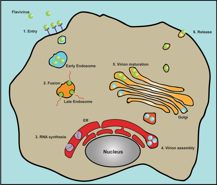
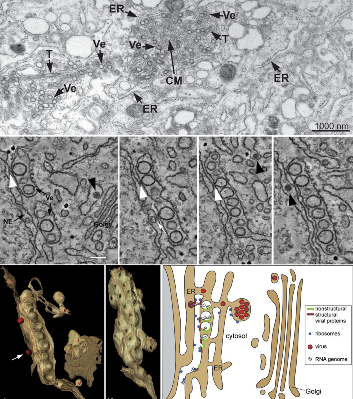
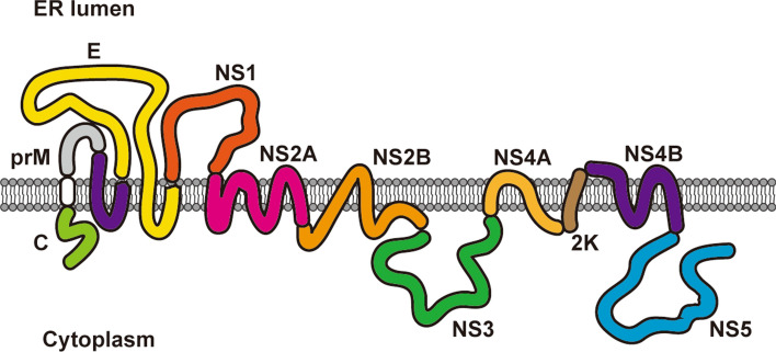
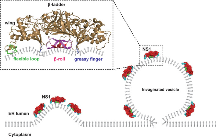
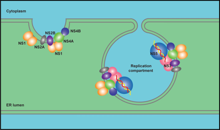
Flaviviruses are positive-sense single-stranded RNA viruses that pose a considerable threat to human health. Flaviviruses replicate in compartmentalized replication organelles derived from the host endoplasmic reticulum (ER). The characteristic architecture of flavivirus replication organelles includes invaginated vesicle packets and convoluted membrane structures. Multiple factors, including both viral proteins and host factors, contribute to the biogenesis of the flavivirus replication organelle. Several viral nonstructural (NS) proteins with membrane activity induce ER rearrangement to build replication compartments, and other NS proteins constitute the replication complexes (RC) in the compartments. Host protein and lipid factors facilitate the formation of replication organelles. The lipid membrane, proteins and viral RNA together form the functional compartmentalized replication organelle, in which the flaviviruses efficiently synthesize viral RNA. Here, we reviewed recent advances in understanding the structure and biogenesis of flavivirus replication organelles, and we further discuss the function of virus NS proteins and related host factors as well as their roles in building the replication organelle.
Keywords: Compartmentalization; ER rearrangement; Flavivirus; Host factors; Membrane remodeling; Nonstructural proteins; Replication organelle.
Conflict of interest statement
The authors declare no conflict of interest.
Figures
Similar articles
-
Flavivirus Replication Organelle Biogenesis in the Endoplasmic Reticulum: Comparison with Other Single-Stranded Positive-Sense RNA Viruses.Int J Mol Sci. 2019 May 11;20(9):2336. doi: 10.3390/ijms20092336. Int J Mol Sci. 2019. PMID: 31083507 Free PMC article. Review.
-
Model System for the Formation of Tick-Borne Encephalitis Virus Replication Compartments without Viral RNA Replication.J Virol. 2019 Aug 28;93(18):e00292-19. doi: 10.1128/JVI.00292-19. Print 2019 Sep 15. J Virol. 2019. PMID: 31243132 Free PMC article.
-
Membrane-Associated Flavivirus Replication Complex-Its Organization and Regulation.Viruses. 2021 Jun 3;13(6):1060. doi: 10.3390/v13061060. Viruses. 2021. PMID: 34205058 Free PMC article. Review.
-
Roles of the Endogenous Lunapark Protein during Flavivirus Replication.Viruses. 2021 Jun 22;13(7):1198. doi: 10.3390/v13071198. Viruses. 2021. PMID: 34206552 Free PMC article.
-
The ACBD3 protein coordinates ER-Golgi contacts to enable productive TBEV infection.J Virol. 2025 May 20;99(5):e0222424. doi: 10.1128/jvi.02224-24. Epub 2025 Apr 10. J Virol. 2025. PMID: 40207930 Free PMC article.
Cited by
-
Papaya Leaf Extracts as Potential Dengue Treatment: An In-Silico Study.Int J Mol Sci. 2022 Oct 14;23(20):12310. doi: 10.3390/ijms232012310. Int J Mol Sci. 2022. PMID: 36293162 Free PMC article.
-
Antiviral plant-derived natural products to combat RNA viruses: Targets throughout the viral life cycle.Lett Appl Microbiol. 2022 Sep;75(3):476-499. doi: 10.1111/lam.13637. Epub 2022 Jan 25. Lett Appl Microbiol. 2022. PMID: 34953146 Free PMC article. Review.
-
Astrocytes derived from neural progenitor cells are susceptible to Zika virus infection.PLoS One. 2023 Mar 29;18(3):e0283429. doi: 10.1371/journal.pone.0283429. eCollection 2023. PLoS One. 2023. PMID: 36989308 Free PMC article.
-
The Japanese encephalitis virus NS1 protein concentrates ER membranes in a cytoskeleton-independent manner to facilitate viral replication.J Virol. 2025 Mar 18;99(3):e0211324. doi: 10.1128/jvi.02113-24. Epub 2025 Feb 5. J Virol. 2025. PMID: 39907281 Free PMC article.
-
Epidemiology, biology, pathogenesis, clinical manifestations, and diagnosis of dengue virus infection, and its trend in Ethiopia: a comprehensive literature review.Trop Med Health. 2023 Feb 24;51(1):11. doi: 10.1186/s41182-023-00504-0. Trop Med Health. 2023. PMID: 36829222 Free PMC article. Review.
References
-
- Bhatt S, Gething PW, Brady OJ, Messina JP, Farlow AW, Moyes CL, Drake JM, Brownstein JS, Hoen AG, Sankoh O, Myers MF, George DB, Jaenisch T, Wint GR, Simmons CP, Scott TW, Farrar JJ, Hay SI. The global distribution and burden of dengue. Nature. 2013;496(7446):504–507. doi: 10.1038/nature12060. - DOI - PMC - PubMed
Publication types
MeSH terms
Substances
Grants and funding
LinkOut - more resources
Full Text Sources
Other Literature Sources
Miscellaneous
