

Angiogenesis imaging study using interim [18F] RGD-K5 PET/CT in patients with lymphoma undergoing chemotherapy: preliminary evidence
- PMID: 33846870
- PMCID: PMC8041962
- DOI: 10.1186/s13550-021-00776-9
Angiogenesis imaging study using interim [18F] RGD-K5 PET/CT in patients with lymphoma undergoing chemotherapy: preliminary evidence
Abstract
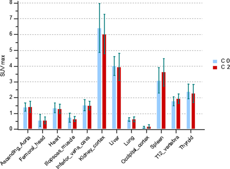
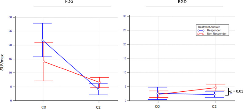
Background: Our aim was to measure the impact of two cycles of standard chemotherapy on tumoural neoangiogenesis by [18F] fluorine arginine-glycine-aspartic (RGD-K5) positron emission tomography-computed tomography (PET) on patients presenting with lymphoma. Nineteen patients at Rouen's Henri Becquerel Cancer Centre were prospectively included. Fluorodeoxyglucose (FDG) and RGD-K5 PET were performed before (C0) and after (C2) two cycles of chemotherapy. End-of-treatment FDG PET was performed for final evaluation. Maximum standardised uptake value (SUVmax), SUVmean, Metabolic Tumour Volume (MTV) and Angiogenic Tumour Volume (ATV) were measured for all lesions. RGD SUVmax and SUVmean were also analysed in 13 normal organs at C0 and C2. The patient's treatment response was considered using the Deauville score (DS) at the end of FDG PET treatment (DS 1-3 were considered responders, and 4 and 5 non-responders).
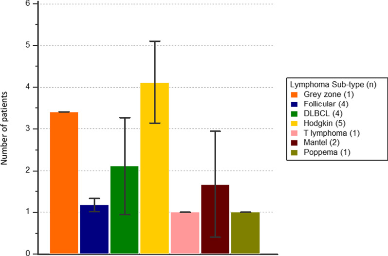
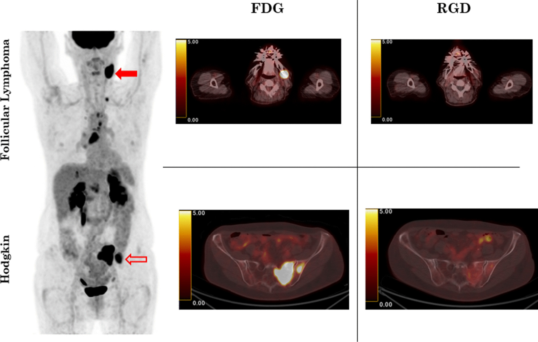
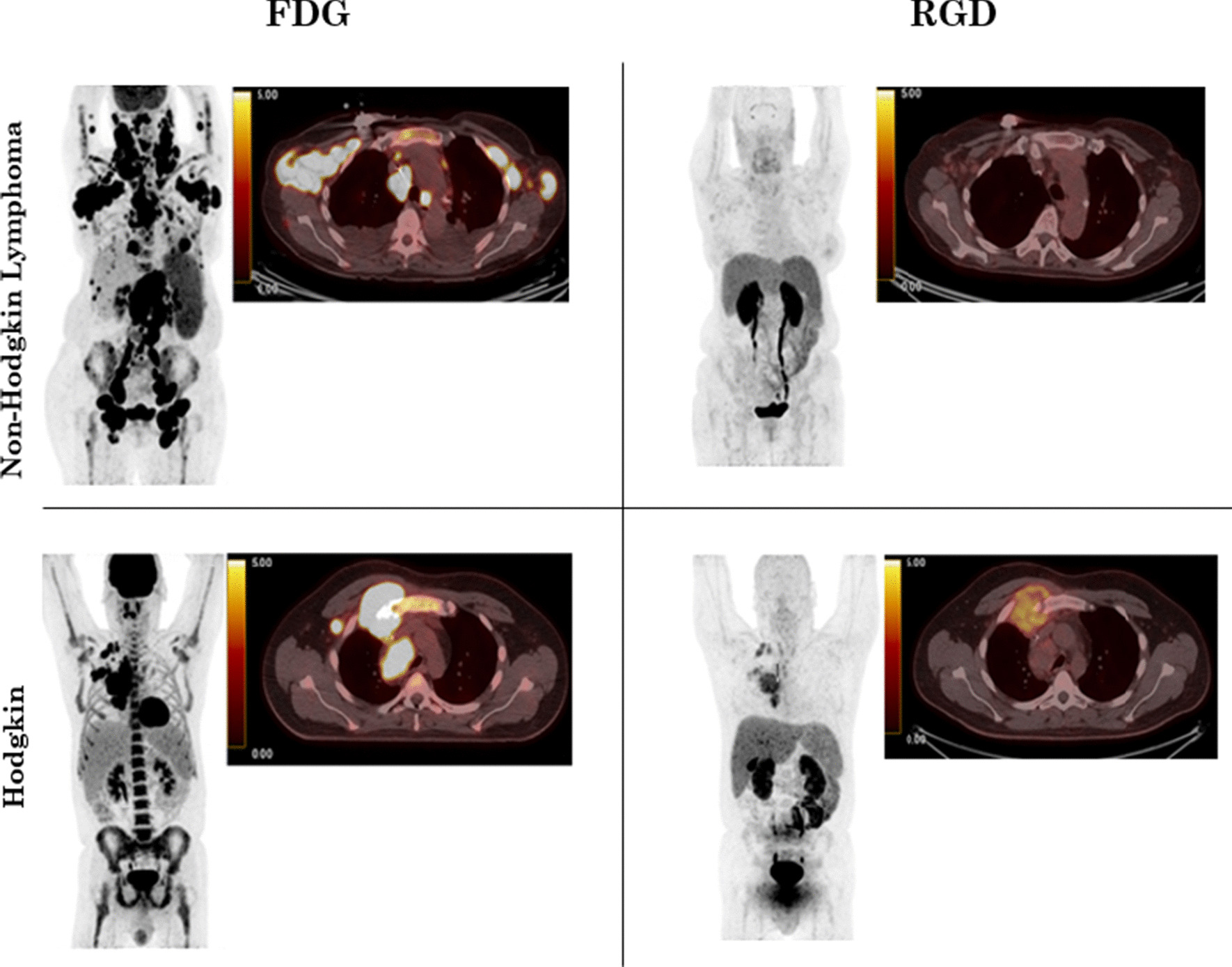
Results: Eighteen patients had both C0 FDG and RGD PET. Twelve patients had both C2 FDG and RGD, completed the treatment protocol and were included in end-of-treatment analysis. No statistical difference was found in RGD uptake of normal organs before and after chemotherapy for SUVmax and SUVmean. On C0 RGD, apart from classical Hodgkin lymphoma (cHL; n = 5) and grey zone lymphoma (GZL; n = 1), other lymphoma sub-types (n = 12) had low RGD uptake (p < 0.001). Regarding FDG, there was no significant difference for SUVmax, SUVmean and MTV at C0 and C2 between patients with cHL and non-Hodgkin lymphoma (NHL). At C2 RGD, non-responders had higher SUVmax and SUVmean compared to responders (p < 0.001). There was no significant difference in RGD ATV between responders and non-responders.
Conclusions: Our study showed significant higher initial RGD uptake in patients presenting with cHL and GZL compared to NHL. Non-responder also had higher post-chemotherapy RGD uptake compared to responders. Issues raised by RGD uptake, particularly in cHL, are yet to be explored and need to be confirmed in a larger population.
Keywords: Angiogenesis; FDG; K5; Lymphoma; PET/CT; RGD.
Conflict of interest statement
All authors declare that they have no competing interests.
Figures
Similar articles
-
(18)F-FPPRGD2 PET/CT: pilot phase evaluation of breast cancer patients.Radiology. 2014 Nov;273(2):549-59. doi: 10.1148/radiol.14140028. Epub 2014 Jul 16. Radiology. 2014. PMID: 25033190
-
The Association Between Liver and Tumor [18F]FDG Uptake in Patients with Diffuse Large B Cell Lymphoma During Chemotherapy.Mol Imaging Biol. 2017 Oct;19(5):787-794. doi: 10.1007/s11307-017-1044-3. Mol Imaging Biol. 2017. PMID: 28144908
-
Predictors of metabolic response in propensity-matched lymphoma patients on interim 18F-fluorodeoxyglucose positron-emission tomography/computed tomography using standardized imaging and reporting protocol: Do we really have one?World J Nucl Med. 2019 Apr-Jun;18(2):154-159. doi: 10.4103/wjnm.WJNM_41_18. World J Nucl Med. 2019. PMID: 31040747 Free PMC article.
-
More advantages in detecting bone and soft tissue metastases from prostate cancer using 18F-PSMA PET/CT.Hell J Nucl Med. 2019 Jan-Apr;22(1):6-9. doi: 10.1967/s002449910952. Epub 2019 Mar 7. Hell J Nucl Med. 2019. PMID: 30843003
-
Whole-body diffusion-weighted MR and FDG-PET/CT in Hodgkin Lymphoma: Predictive role before treatment and early assessment after two courses of ABVD.Eur J Radiol. 2018 Jun;103:90-98. doi: 10.1016/j.ejrad.2018.04.014. Epub 2018 Apr 17. Eur J Radiol. 2018. PMID: 29803392
Cited by
-
Advance in peptide-based drug development: delivery platforms, therapeutics and vaccines.Signal Transduct Target Ther. 2025 Mar 5;10(1):74. doi: 10.1038/s41392-024-02107-5. Signal Transduct Target Ther. 2025. PMID: 40038239 Free PMC article. Review.
-
Tumor angiogenesis at baseline identified by 18F-Alfatide II PET/CT may predict survival among patients with locally advanced non-small cell lung cancer treated with concurrent chemoradiotherapy.J Transl Med. 2022 Feb 2;20(1):63. doi: 10.1186/s12967-022-03256-3. J Transl Med. 2022. PMID: 35109866 Free PMC article. Clinical Trial.
-
Peptide-drug conjugates (PDCs): a novel trend of research and development on targeted therapy, hype or hope?Acta Pharm Sin B. 2023 Feb;13(2):498-516. doi: 10.1016/j.apsb.2022.07.020. Epub 2022 Aug 3. Acta Pharm Sin B. 2023. PMID: 36873165 Free PMC article. Review.
References
LinkOut - more resources
Full Text Sources
Other Literature Sources
Research Materials
Miscellaneous
