

Excision of fibrous band and application of tension band plating in focal fibrocartilaginous dysplasia: A case report and literature review
- PMID: 33847584
- PMCID: PMC11229613
- DOI: 10.5152/j.aott.2021.20080
Excision of fibrous band and application of tension band plating in focal fibrocartilaginous dysplasia: A case report and literature review
Abstract
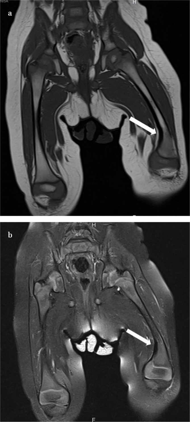
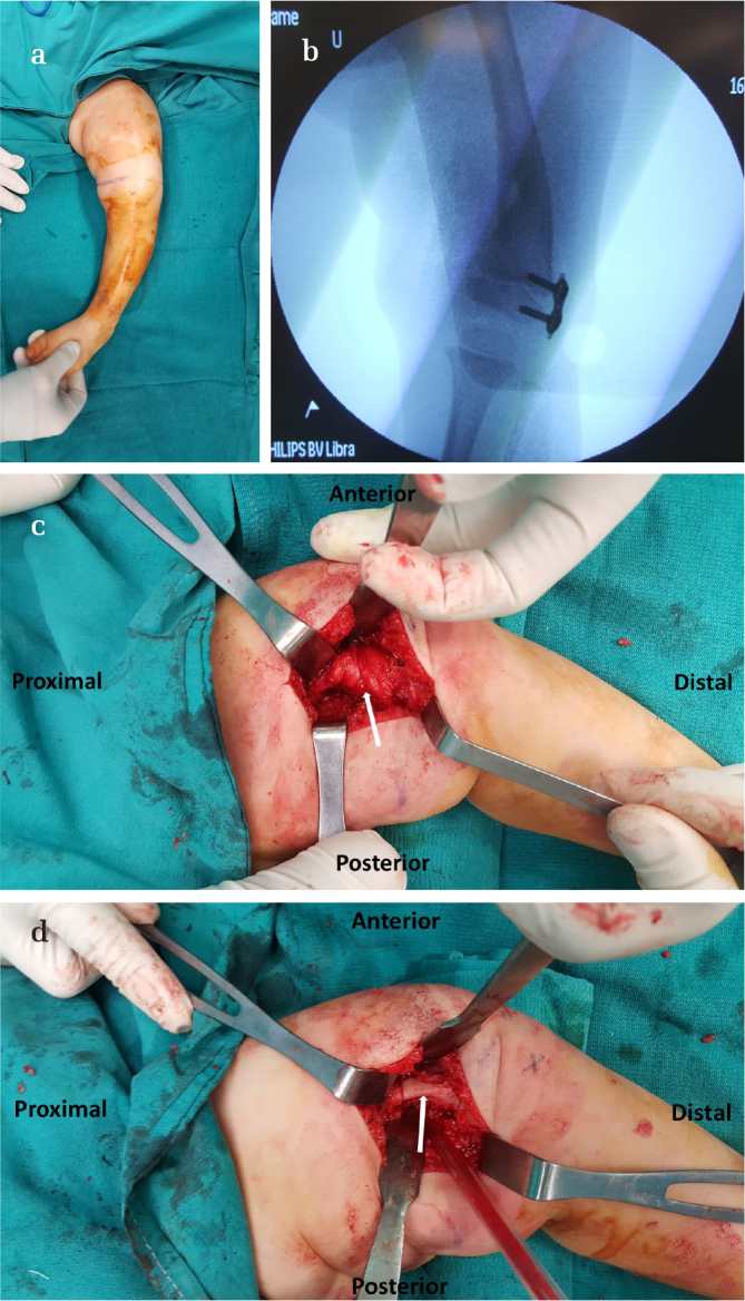
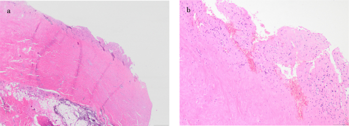
Focal fibrocartilaginous dysplasia (FFCD) is a rare disease that can cause angular deformities of long bones. The common pathologic finding is a thick fibrotic band extending from epiphysis to metaphysis on one side of the bone. The tethering effect of the fibrotic band around the growth plate is thought to be the main etiology for the development and progression of the deformity. FFCD mostly affects the proximal tibia and the distal femur. The literature contains different treatment options. Here, we present the case of a 20-month-old girl with FFCD on the medial side of the distal femur causing varus deformity. Our treatment protocol included excision of the fibrotic band from the medial side and application of a two-hole plate for guided growth on the lateral side of the distal femur. Deformity correction was achieved rapidly with no complications. A literature review is also presented along with pathologic and magnetic resonance imaging findings.
Conflict of interest statement
Figures

Similar articles
-
Spontaneous resolution of angular deformity of the distal femur in focal fibrocartilaginous dysplasia: a case report.J Pediatr Orthop B. 2010 Mar;19(2):161-3. doi: 10.1097/BPB.0b013e3283361b11. J Pediatr Orthop B. 2010. PMID: 20051915
-
Correction of Angular Deformities Due to Focal Fibrocartilaginous Dysplasia Using Guided Growth: A Preliminary Report.J Pediatr Orthop. 2017 Apr/May;37(3):e183-e187. doi: 10.1097/BPO.0000000000000785. J Pediatr Orthop. 2017. PMID: 27261964 Review.
-
Tibia valga due to focal fibrocartilaginous dysplasia: case report.J Pediatr Orthop B. 2002 Apr;11(2):167-71. doi: 10.1097/00009957-200204000-00015. J Pediatr Orthop B. 2002. PMID: 11943993 Review.
-
A patient with focal fibrocartilaginous dysplasia in the distal femur and review of the literature.Tohoku J Exp Med. 2008 Aug;215(4):307-12. doi: 10.1620/tjem.215.307. Tohoku J Exp Med. 2008. PMID: 18679004
-
Focal fibrocartilaginous dysplasia of the tibia: long-term evolution.Acta Orthop Belg. 2006 Jan;72(1):77-82. Acta Orthop Belg. 2006. PMID: 16570899
Cited by
-
Fibrocartilaginous dysplasia: What do we know so far?Radiol Case Rep. 2023 Feb 28;18(5):1763-1766. doi: 10.1016/j.radcr.2023.01.074. eCollection 2023 May. Radiol Case Rep. 2023. PMID: 36926535 Free PMC article.
References
-
- Müezzinoğlu B, Öztop F. Fibrocartilaginous dysplasia: a variant of fibrous dysplasia. Malays J Pathol. 2001;23:35–40. - PubMed
Publication types
MeSH terms
LinkOut - more resources
Full Text Sources
Other Literature Sources
