

Postmortem coronary artery calcium score in cases of myocardial infarction
- PMID: 33847801
- PMCID: PMC8354952
- DOI: 10.1007/s00414-021-02586-z
Postmortem coronary artery calcium score in cases of myocardial infarction
Erratum in
-
Correction to: Postmortem coronary artery calcium score in cases of myocardial infarction.Int J Legal Med. 2021 Sep;135(5):2141. doi: 10.1007/s00414-021-02608-w. Int J Legal Med. 2021. PMID: 34021397 Free PMC article. No abstract available.
Abstract
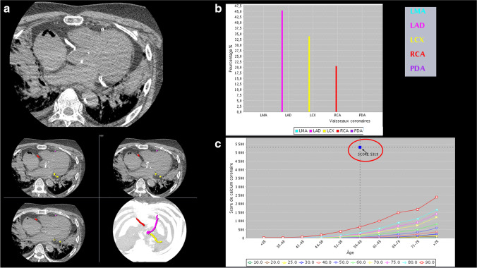
Sudden cardiac death (SCD) related to atherosclerotic coronary artery disease (ACAD) resulting in myocardial infarction is the most prevalent cause of death in western countries. In clinical practice, coronary artery calcium score (CACS) is considered an independent predictor of coronary events, closely related to atherosclerotic burden and is quantified radiologically by the Agatston score being calculated through computed tomography. Postmortem computed tomography (PMCT) allows the visualization and quantification of coronary calcifications before the autopsy. However, it was reported that some patients who died from severe ACAD had a zero CACS in PMCT. In this study, a retrospective evaluation of CACS in adult's myocardial infarction cases related to ACAD, with available CACS and histological slides of coronary arteries, was performed in order to gain a deeper understanding of coronary calcifications and their role in myocardial infarction cases. The CACS was calculated by using the software Smartscore 4.0 after the radiological examination on a 64-row CT unit using a specific cardiac protocol. Thirty-six cases were identified out of 582 autopsies, recorded during a 2-year study period (29 men, 7 women; age 56.3 ± 11.7). CACS was 0-10 in 5 cases (5 men, 44.8 ± 13.7), 11-100 in 8 cases (6 men, 2 women, 53.1 ± 7.7), 101-400 in 13 cases (11 men, 2 women, 57.4 ± 9.6), and > 400 in 10 cases (9 men, 1 woman, 63.1 ± 11.9). Coronary thrombosis was found in 28 cases, histologically identified as plaque erosions in 6 cases and as plaque ruptures in 22 cases. Statistical analyses showed that CACS increases significantly with age (p-value < 0.05) and does not show significant correlation with gender, body weight, body mass index, and heart weight. CACS was significantly higher in plaque ruptures than in plaque erosions (p-value < 0.01). Zero or low CACS on unenhanced PMCT cannot exclude the presence of myocardial infarction related to ACAD. This paradoxical discrepancy between imaging and autopsy findings can be explained considering the histological aspect of fatal coronary plaques.
Keywords: Coronary artery calcium score; Coronary calcifications; Postmortem imaging; Sudden cardiac death.
© 2021. The Author(s).
Conflict of interest statement
This study is in accordance with the Swiss ethical standards.
The authors declare no competing interests.
Figures


References
-
- Basso C, Aguilera B, Banner J, Cohle S, d'Amati G, de Gouveia RH, di Gioia C, et al. Guidelines for autopsy investigation of sudden cardiac death: 2017 update from the Association for European Cardiovascular Pathology. Virchows Arch. 2017;471(6):691–705. doi: 10.1007/s00428-017-2221-0. - DOI - PMC - PubMed
MeSH terms
LinkOut - more resources
Full Text Sources
Other Literature Sources
Medical
